Blog

  • ACL Injury

    Knee injuries, especially the anterior cruciate ligament (ACL)  injuries, are a potentially devastating problem among athletes and impose substantial time loss in a player’s career and financial burdens on athletes’ families and the healthcare system. In this blog we intend inform about exercises for ACL recovery as well.

    ACL INJURY STATISTICS:-

    This study of level 4 evidence conducted on Indian athletes revealed that ACL injuries compromised 86.5% of all the knee injuries. (1)

    WHAT IS ACL INJURY?

    ACL is one of the central knee ligaments which is responsible for maintaining the knee stable during various activities. The ligament may be completely torn, partially torn, or avulsed from its origin or insertion. The ACL is the primary restraint to excessive anterior translation and rotation of the tibia on the femur. (4) It is one of the most common injuries worldwide, which usually may occur along with other structures in the knee.

    CAUSES OF ACL INJURY

    This injury may occur while playing sports like football, rugby, cricket, kabaddi, basketball, and handball or may occur due to trauma. The incidence rate is higher in women compared to men, the reason being wider pelvis, hormonal changes, and weak neuromuscular co-ordination issues. (1) Complete ACL disruption typically results in dynamic knee instability or the inability to respond to quick changes in position. (4)

    ACL ligament exercises are usually based on all these points which are guided by our expert physiotherapist. we recommend you to sign up for physio for ACL tear with us for the best results

    ACL INJURY MECHANISM:-

    Figure 1: Tackled by another player leading to vagus load

    The mechanism by which an ACL injury or a tear can occur can be traumatic or non-traumatic. Nontraumatic mechanism is faulty landing from a jump, sudden turns, and sudden stops while sprinting. Traumatic could be tackled from fellow players or accidents.

                                                                 Figure 2 Faulty jump landing.

    other than the mechanisms, it is important to look into Movement factors increasing susceptibility to ACL Injuries.

    (more…)

  • Acute Injury Management

    Musculoskeletal or sports injuries are injuries that happen when playing sports or exercising.

    There are two kinds of injuries:

    acute injury mgmt

    • Chronic injuries happen after you play a sport or exercise for a long time.

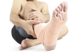
    The first 24-48 hours after sustaining a soft tissue injury are crucial in ensuring the best outcome for healing.  As a general rule, management for most injuries is as simple as remembering two simple acronyms – RICER and HARM.

    The objective of RICE is to stop the injury-induced bleeding into the muscle tissue and thereby minimize the extent of the injury. Clinical examination should be carried out immediately after the injury and 5-7 days after the initial trauma, at which point the severity of the injury can be assessed more reliably.

    Use RICER!

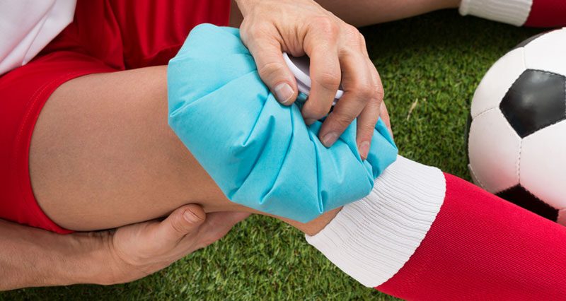
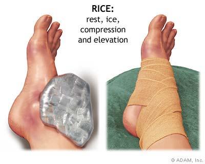
    RICER stands for Rest, Ice, Compression, Elevation, and Refer. RICER highlights 5 simple methods to reduce pain and promote healing.

    Rest – Initially, the objective for healing any soft tissue injury is to minimise swelling and bleeding. Reducing movement, and in some cases immobilising the injured area, is one of the simplest methods to reduce further injury and promote healing 6.

    Ice – Cooling the injured area serves to slow down tissue metabolism 1,2,4. It may cause contraction of blood vessels which will slow down blood flow and assist with reducing swelling 1,2,4. Ice can also provide a temporary numbing effect to assist with short-term pain relief as well as 1,2,4. Ensure that intense cooling is not applied directly to the skin to minimize the potential for skin burns. Applying ice following common injuries such as Rotator cuff tear, Hamstring Strain or Ankle Sprains is usually a good start.

    Compression – Compressing an injured area can have a direct effect on swelling. Applying a bandage to the area can help to limit the amount of bleeding that occurs in an isolated area 3. Caution needs to be taken to ensure that any compressive bandage is firm, however, does not reduce the normal circulation to the area.

    Elevation – Elevating the injured area will also serve to reduce the collection of fluid to the area by limiting the effect of gravity 6. For foot or leg injuries, lying with the injured area elevated above the level of the pelvis with the use of pillows is an easy start.

    Refer – While RICER helps promote healing in the early stages of injury to get the right diagnosis you need to see an appropriate health professional; say a physiotherapist! Here at Physiocure, we can give you an accurate diagnosis and also help plan a return to the sporting field.

     Usually, the treatment of injured muscles should be carried out by immediate immobilization of the injured muscle (clinically, relative immobility/avoidance of muscle contractions). However, the duration of immobilization should be limited to a period sufficient to produce a scar of sufficient strength to bear the forces induced by remobilization without re-rupture and the return to activity (mobilization) should then be started gradually within the limits of pain.

    At Physiocure we aim at, early return to activity, which is needed to optimize the regeneration of healing muscle and recovery of the flexibility and strength of the injured skeletal muscle to pre-injury levels. Our rehab programs are built around progressive agility and trunk stabilization exercises, as these exercises seem to yield a better outcome for injured skeletal muscle than programmes based exclusively on stretching and strengthening of the injured muscle.

    Braces

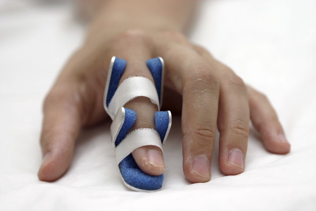
    Braces and splints can be useful for acute injuries, chronic conditions, and the prevention of injury. There is good evidence to support the use of some braces and splints; others are used because of subjective reports from patients, relatively low cost, and few adverse effects, despite limited data on their effectiveness
    They can be used to immobilize an unstable joint or fracture, to unload a portion of a joint and improve pain and function, to eliminate range of motion in one direction, or to modify range of motion in one or more directions. They do not replace a good rehabilitative program, and the entire spectrum of treatment options should be explored and used as needed.

    For acute injury or during a post-operative period, common braces include:
    • walking cast
    • wrist splint
    • ankle brace
    • knee brace
    • finger/toe splints
    • casts (hard and removable)

    An accurate diagnosis of the injury is important in determining whether a brace or splint is indicated. Generally, splints are for short-term use. Excessive, continuous use of a brace or splint can lead to chronic pain and stiffness of a joint or to muscle weakness. However, long-term use of some braces, such as a knee unloader brace, can help prevent the progression of pain attributable to osteoarthritis of the knee.

    Avoid HARM!

    HARM stands for Heat, Alcohol, Running/ exercise,and Massage. HARM represents 4 factors that you should avoid in the early stages of injury in order to maximise healing and recovery. While RICER focuses on minimising bleeding and swelling, HARM covers factors to avoid – all of which will increase circulation to the damaged area.

    Heat – Heat will cause blood vessels to dilate which in turn will increase the flow of blood to the area. Avoid hot baths, showers, saunas, heat packs, and heat rubs.

    Alcohol – Similar to heat, alcohol has an effect of dilating blood vessels, which in turn will increase the flow of blood to the area. Alcohol can also mask pain and the severity of the injury, which may put you at greater risk for re-injury 7.The alcohol effects on sports performance depend on the type and dosage of alcohol, acute vs chronic administration, the alcohol elimination rate as well as the type of exercise. Avoid drinking alcohol in the initial stages of healing any injury.

    Running/exercise – An increase in heart rate increases the flow of blood around the body. This will cause blood to accumulate in the area faster. Take the opportunity to rest.

    Massage – Massaging the area, once again, will stimulate the flow of blood to the area. Avoid massage in the initial stages of injury.

    References:

    1.  The use of ice in the treatment of acute soft-tissue injury.
    2. Cryotherapy for acute ankle sprains: a randomised controlled study of two different icing protocols.
    3. Brukner B, Khan K. Clinical Sports Medicine: Third Edition, McGraw Hill, Australia Pty, Ltd. 2007.
    4. Muscle injuries: optimising recovery. 
    5. The effect of exercise, alcohol or both combined on health and physical performance.
    6. Braces and Splints for Musculoskeletal Conditions


  • Squats During Pregnancy

    Each woman has a different take on pregnancy based on the lifestyle, culture, general fitness, complications accompanied with the pregnancy. In this article we want to show research based benefits of squats during a “HEALTHY PREGNANCY”. We strictly advise your to consult a gynaecologist and your ANC consultant before you try these exercises.

    Research shows that exercise during pregnancy has a positive effect on the pregnancy and labour. Similarly performing squats throughout the pregnancy can have following benefits:-

    1. Keeps Lower back, core muscles and lower body strength maintained.
    2. Helps in opening up hips and pelvis by the third trimester , which helps in easier labour.
    3. Helps in maintaining the strength of the pelvic floor muscles
    4. Can prevent back pain during and after pregnancy.
    5. Helps in maintaining a good posture during pregnancy.
    6. Prevents excessive weight gain

    v

    In the past few years a lot of pregnant ladies who came in to learn ANC exercises expressed concerns like, performing squats causes increased intra-abdominal pressure, which affects the blood supply to the baby. The second myth I came across is avoid performing full squats or for that matter bending forwards or sitting on the ground as it may affect the baby’s growth. As an ANC consultant , I would say it differs from case to case.

    Given the vast Indian cultural history, our ancestors have given births without any science to support them. women were working in the kitchen or the house with the need to squat or sit on the ground or a small step for every little chore. By just doing their everyday chores while sitting on the ground and getting up  they squatted several number of times in a day and still normal babies where born. So its absurd not to sit on the ground or not to squat.

    Secondly if considered squats as a form of resistance exercises there is research to prove squats do not affect the pregnancy or the baby health. Check out this research paper if you are into fact finding spree

    https://obgyn.onlinelibrary.wiley.com/doi/pdf/10.1111/aogs.12525

    The take away:- well this research doesn’t about safety of squats but it says resistance exercises in general lead to a better out come. The sample size is small, but some evidence is better than none.

    Deep squatting seems to offer a better overall way to stretch and align the pelvis in pregnancy and help the baby to descend.  Many of the fifty or so clips and articles I’ve reviewed wisely , but only a few of the web articles differentiate between the parallel and deep squat and even less listed are any contra indications.

    The contraindications to doing deep squats are:-

    1. hemorrhoids,
    2. vasa previa,
    3. placenta previa or
    4. a breech lie.

    But perhaps the more serious contraindication for squats is lightening (when the baby descends into the upper pelvis) which normally occurs around the 34-38 week mark in primips but could be earlier or later

    Biomechanically seeing the full squat, it has its pros and cons. If done with the correct technique, it has the above mentioned benefits. But when done a full squat in a wrong manner can put undue pressure on the pubic joints or the sacroiliac joints in your pelvic girdle. So make sure you learn the correct form from your certified trainer/yogi/physio if you are inexperienced in performing a full squat.

    Here is a list of different type of squats which you can do safely during pregnancy

    (1)BODY WEIGHT SQUATS

    FULL/ DEEP SQUATS

    During pregnancy, the weight of your body may offer enough resistance for you to work out effectively. But you can always add weight by holding dumbbells in each hand, or by setting a barbell across your shoulders.

    1. Stand with feet shoulder-width apart.
    2. Hold your arms straight out in front of your body for balance if you don’t have weights or a bar.
    3. Lower yourself into a full or a half squat position. Only go as far as you’re comfortable while keeping your back straight, weight in your heels, and knees behind or in line with your toes.
    4. Return to starting position, squeezing your glutes on the way up.
    5. Perform 3 sets of 10 to 15 repetitions.
    HALF/PARELLEL SQUATS

    Note :-

    1. performing full squats in the right form aides the above mentioned benefits
    2. Avoid the half squats if you have never worked out before or you have existing knee pain.
    3. Performing the squats in front of the mirror, helps you in achieving the correct form.
    4. Weights or resistance to be added only after consulting your personal trainer and/or physio.

    (2) SUMO SQUATS

    This squat variation targets the inner muscles of the thighs and glutes. It’s also an excellent stretch to open up the hips.

    Note: Your joints are more flexible during pregnancy, so it’s easy to strain yourself by stretching too far. Do not go past your normal range of motion.

    1. Step out into a wide stance with your feet greater than shoulder-width apart and toes pointing outward, with knees tracking in line with the toes.
    2. Lower yourself into a squat position. Only go as far as you’re comfortable while keeping your back straight, weight in your heels, and knees behind or in line with your toes.
    3. Keep your legs turned out throughout the movement, making sure your knees do not cave in toward each other.
    4. Return to the starting position, squeezing your glutes on the way up.
    5. Perform 3 sets of 10 to 15 repetitions.


    1. SQUATS AGAINST WALL WITH EXERCISE BALL

    This exercise adds a level of instability to further engage the core muscles during the squat movement. If this exercise bothers your knees, only go as low as you feel comfortable.

    1. Stand against a wall with an exercise ball between the wall and your lower back.
    2. Place feet shoulder-width apart.
    3. Hold your arms straight out in front of your body for balance.
    4. Contract your abdominals by pulling in your bellybutton as if you were trying to pull it to the ball behind you.
    5. Lower yourself into a sitting position. Only go as far as you’re comfortable while keeping your back straight and shoulders back.
    6. If you find you have a lot of pressure on your knees, make sure your feet are far enough away from the wall to maintain a 90-degree angle at the knee when in a full squat.
    7. Return to starting position, squeezing your glutes on the way up.
    8. Perform 3 sets of 10 to 15 repetitions.


  • Overhead Squat Assesment

    Are you a sports professional, an athlete, or a fitness freak, do you succumb to lower-body injuries very easily or do you want to correct your body alignment???

    How can a squat assessment help you?

    Basically, a squat assessment can help us gauge

    • the possibility of deficiency in mobility in the hip, knee, or ankle joints.
    • secondly, it helps us identify muscle weaknesses in the lower back, hip, or ankle which have a scope of improvement.
    • thirdly it could be contributing to an injury that would develop on practicing a wrong form of squat.
    • lastly, you can get it done to get the form right from the beginning itself or if you are gyming after an injury or surgery in the lowerbody.

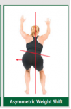
    squat could look like this when observed from behind but you may not be able to

    asymmetric squats
    asymmetric squats

    understand the faults. this fault is very common with individuals recovering from a knee injury or a knee surgery, so as an experienced physio I would advise you to get the assessment done for activating the correct neuromuscular pattern during the squat

    Just one simple test of Over Head Squat Assessment (OHSA) can highlight the shortcomings in your form and help you perform better.

    The ability to squat is essential in life whether you are a sports person or just a regular person. You need to squat for your daily activities right from the age of 2 to 80 years old. A lot of patients come to us not knowing how to squat because of lifestyle issues, on the other hand, a lot of sports players, who come in for treatments have shown asymmetrical strength and weaknesses, we could identify through this simple test to help then recover completely from their back/hip/knee/ankle injury.

    When you are squatting, all the muscles in the linkage right from your toe to the spine to the shoulder and neck muscles must work in co-ordination and appropriately to produce an effective squat.

    An OHSA requires you to perform squats for around 5-10 reps and the form of your squats are assessed through 3 different angles

    if there are any faults in performing the squats, the same are identified and a correction program is made to help you perform better.

    faulty squat pattern- front view


    faulty squat pattern- side view

    fill in the OHSA form and come over to get yourself corrected. if you cannot we can do this assessment

    for you.

    PS:- OHSA is not the only tool, it forms a part of a group of the examination for various musculoskeletal/sports injuries.

    Overhead Squat Assesment form

  • Effects of Exercises in Pregnancy

    For long now, women have been scared away from exercising during pregnancy, but there are more and more articles floating on the web talking about all the positive effects of exercising during a normal pregnancy. At Physiocure, we advise all women with a normal pregnancy to adopt some form of safe prenatal exercises to have an even better and injury-free time, once their baby arrives.


    Current studies indicate that healthy women with an uncomplicated pregnancy do not need to limit their exercise for fear of adverse effects (Whaley et al. 2006). Women should be encouraged to do consistent, moderate-intensity exercise, however women with complicated pregnancies have been discouraged from participating in exercise activities for the fear of impacting the underlying disorder (Davies et al. 2003). According to Kramer & McDonald (2009) regular aerobic exercise during pregnancy appears to improve physical fitness. Women who are exercising prior to the pregnancy can continue to do so without any major modifications. However, many women will change their program because they have symptoms, discomfort and risks associated with joint laxity and body mass gain (Whaley et al. 2006). This joint laxity should also be taken in consideration during stretching. Most woman start exercise programs in the second trimester because often the symptoms as nausea, vomiting and profound fatigue are less in that stage.

    Exercise prescription

    During pregnancy maximal exercise testing is not recommended. If a test is needed this should be undertaken under the supervision of a physiotherapist. Women who were less active or sedentary before the pregnancy should go to a physiotherapist before they start exercising.The recommended mode, frequency, duration and overload of the exercise are consistent with general principles of fitness training (Table 1).

    Table 1: GENERAL FITNESS EXERCISE PROGRAM (Whaley 2006)
    Components Frequency Intensity Duration Activity
    Cardio respiratory 3-5 /week 40% – 85% HRR or Vo2R 55% – 90% HRmax   12-16 RPE 20-60 min Large muscle groups

    Dynamic activity

    Resistance 2-3 /week Volitional fatigue (MMF) (e.g. 19-20 RPE)

    OR

    Stop 2-3 reps before volitional fatigue (e.g. 16 RPE)

    1 set of 3-20 repetitions (e.g. 3-5, 8-10, 12-15) 8-10 exercises IncRemove featured imagelude all major muscle groups
    Flexibility Minimal

    2-3/ week

    Ideally 5-7/week

    Stretch to tightness at the end of the range of motion but not to pain 15-30 seconds  2-4 repititions Static stretch all major muscle groups

    Research also reports that light resistance training can be performed over the 2nd and 3rd semester and that does not have an effect on the newborn body size or overall health. They conclude that resistance training during pregnancy decreases the negative effects of maternal weight on the newborn size. Davies et al. (2003) recommends the `talk test´ method to monitor exertion, which means that the woman must be able to maintain a conversation during exercise. He explains that if she is unable to do so, reduced exercise is advised.

    Table 2: MODIFIED HEART RATE TARGET ZONES FOR AEROBIC EXERCISE IN PREGNANCY (Davies 2003)
    Maternal Age ( in years) Heart Rate Target Zone       

            (beats/min)

    Heart Rate Target Zone

           (beats/10 sec)

    Less than 20 140 – 155 23 – 26
    20-29 135 – 150 22 – 25
    30-39 130 – 145 21 – 24
    40 or greater 125 – 140 20 – 23


    According to ACOG (2002) a prescription of 30 to 40 minutes or more of moderate activity on most or all of the days in the week is advised and 3-5 times a week for more intense physical activities. There is a difference between women who have been inactive prior to pregnancy and women who have been active. The inactive women should begin with a light-intensity program (20%-39% HRR), this means low impact activities such as walking and swimming. Previously sedentary woman should start with 15 minutes exercise, 3 times a week, increasing this gradually to 30 minutes session, four times a week. He recommends that in this session abdominal strengthening should be excluded as there may be development of diastasis recti and associated muscle weakness. However core stability is an important part of exercise during the pregnancy. Goals should include maintenance of cardiorespiratory fitness, muscle mass and physician-recommended weight gain (McArdle et al. 2006).

    Pelvic floor training>

    Pregnancy and pelvic pain have an important impact on women on a daily basis. Stuge et al. (2002) explains that lumbo-pelvic stabilization could be achieved by exercises and have an influence on appropriate posture and enhanced muscle function or by use of an orthotic device like a pelvic band could have an influence on it. He explains that there is no strong evidence that the effect of physical therapy interventions helps with the prevention of low-back pain and pelvic pain related to pregnancy but there are no facts that exercise doesn’t help. However, he mentions that water gymnastics has shown a lower pain intensity and sick leave compared to no intervention, but this could be due to the effect of attention. A training program for the pelvic floor muscle can be associated with fewer cases of active pushing in the second stage of labour lasting longer than 60 minutes.

    Exercise in the Postpartum Period

    Depending on the type of delivery, most types of exercises can be continued. The women may need to reduce the activities due to fatigue of delivery and newborn care. Brukhner & Khan (2007)  state that the changes caused by pregnancy take some time to return to normal, so care should be taken in the first six weeks after delivery to avoid sudden high impact or contact activity. They explain that if the delivery was a caesarean section, women may need to avoid strenuous activity for six weeks and heavy weight training for 12 weeks. This depends on the recovery such as anemia or wound infection.  Pelvic floor exercises are good to reduce the risk of future urinary incontinence (Morkved & Bo 2000).

    Breastfeeding is a part that also needs to be considered after pregnancy. The exercises should not interfere with the ability to breastfeed. Although exercise doesn’t affect the milk production or composition, lactic acid has been shown to be increased in the breast milk of women exercising at maximal intensity, but not in those who exercising at normal level (Davies et al). 2003) the growth of breastfeeding babies of mothers who exercise is normal. This is even the case for mothers who are losing weight as part of their exercise.


    Exercise to prevent complications

    1.    Gestational diabetes mellitus (GDM)

    Avery (1997) concluded that in women who started a home-based exercise program there was no reduction in glucose levels. According to Dempsey et al. (2004) an increase in physical activity may reduce the risk of gestational diabetes mellitus. Physical activity, especially vigorous activity before pregnancy and at least light to- moderate activity during pregnancy may reduce risk for abnormal glucose tolerance and GDM (Oken et al. 2006). There is inconsistence evidence whether exercise has a positive or negative effect on gestational diabetes.

    2.    Preeclampsia

    Preeclampsia is identified by increased blood pressure and protein in the urine, women usually don’t have any symptoms in the beginning.

    Women who are doing leisure-time exercise during pregnancy where less likely to develop preeclampsia compared to those who didn’t participate in exercise. Relative to inactive women, the pregnant women who were vigorously active during pregnancy experienced a 54% reduction of risk for preeclampsia, and those who engaged in light or moderate activities experienced a 24% reduction of risk (Lewis et al. 2008).


    3.    Weight gain

    Total weight gain is measured in the following categories: low (<10 kg), medium (10–15 kg), high (16–19 kg), or very high (>20 kg). Research explains that generally low gestational weight gain was an advantage for the mother, but it increases the risk of having a small baby, particularly for underweight women. In conclusion heavier women may benefit from avoiding high and very high pregnancy weight gain, which brings only a slight increase in the risk of growth restriction for the infant. High weight gain in underweight women does not appear to have deleterious consequences for them or their infants, but they may want to avoid low gestational weight gain to prevent having a small baby.

    4.    Labor and Birth

    There is an overall positive relationship between a more favourable labor and delivery outcomes and exercise. In addition, even among women with diseases and previous poor outcomes, exercise during the pregnancy was not associated with either preterm birth or low birth weight. Juhl et al. (2007) results show a reduced risk in preterm birth among women who participated in exercise with 40% in comparison with non-exercisers, but no dose-response relation was seen.

    5.   Urinary incontinence

    About one third of the women have an incontinence leakage after they gave childbirth . Training of the pelvic floor muscle is an often recommended treatment during and after childbirth for prevention of incontinence and there is some evidence that pelvic floor muscle training in women having their first baby can prevent urinary incontinence in late pregnancy and postpartum (Hay-Smith et al. 2009). However, Ewings et al. (2005) states that pelvic exercise did not help prevent future incontinence, although this could be due to a failure of the women to return for the classes

    CONCLUSION:-

    In conclusion evidence indicates that exercise during pregnancy is recommended.  An important part of exercise during pregnancy is safety. Therefore listening to their body is a main factor. And pregnant women must be watched carefully and be made aware of conditions such as vaginal bleeding, dyspnea prior to exertion, dizziness, heachache, chest pain, muscle weakness, calf pain or swelling preterm labor, decreased fetal movement and amniotic fluid leakage.

    During exercise the heart rate should be measured closely and the talk test is an easy way to know if the woman is at the correct intensity level. The supine position should be avoided after 4 weeks of pregnancy.

    Finally, your physiotherapist should be well informed about exercise during pregnancy so as to empower you with the ability to have a healthy, safe and enjoyable pregnancy.

    SIMILAR ARTICLES:-

    Squats during pregnancy

    Safety Issues in Pregnancy

    Low Back Pain

    Post Natal Back Pain- a personal take!!

  • Safe Exercises In Preganacy

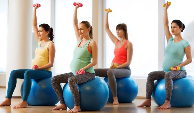
    Safely exercising during pregnancy has more perks than dangers. I have mentioned in almost all my pre and postnatal articles, that when u are able to exercise throughout your pregnancy, it prepares your body well for childbirth as well as for postpartum issues you may face. In this article, I will be covering a range of Do’s and Dont’s in terms during pregnancy to exercise safely.

    ACTIVITIES TO AVOID:-

    Activities that do not impact your and your baby’s health should only be taken up. For instance, Avoid activities where you could take a hard fall. You may have safely ridden a bike in the first trimester, but why risk it now? If bike riding is a critical part of your exercise routine, choose a stationary bike from here on out.

    If you’re an avid skier, stick to the bunny slope, or switch to cross-country. Anything that reduces potential oxygen flow, like scuba diving or activities at high elevation, isn’t safe.

    You should stop exercising if you:

    • feel queasy
    • get too hot
    • feel dehydrated
    • experience any vaginal discharge, bleeding, or abdominal or pelvic pain

    SYMPTOMS TO STOP EXERCISING DURING PREGNANCY

    • Vaginal bleeding
    • Dyspnea prior to exertion
    • Dizziness
    • Heachache
    • Chest pain
    • Muscle weakness
    • Calf pain or swelling (need to rule out thrombophlebitis)
    • Preterm labor
    • Decreased fetal movement
    • Amniotic fluid leakage


    CONTRAINDICATIONS FOR EXERCISES DURING PREGNANCY:-

    Relative Absolute
    ·       Severe trauma

    ·       Unevaluated maternal cardiac dysrhythma

    ·       Chronic bronchitis

    ·       Poorly controlled type I diabetes

    ·       Extreme morbid obesity

    ·       Extreme underweight (BMI<12)

    ·       History of extremely sedentary lifestyle

    ·       Intrauterine growth restriction in current pregnancy

    ·       Poorly controlled hypertension

    ·       Orthopedic limitations

    ·       Poorly controlled seizure disorder

    ·       Poorly controlled hyperthyroidism

    ·       Heavy smoker

    ·       Hemodynamically significant heart disease

    ·       Restrictive lung disease

    ·       Incompetent cervic/cerclage

    ·       Multiple gestation at risk for premature labor

    ·       Persistent second- or third-trimester bleeding

    ·       Placenta previa after 26 weeks of gestation

    ·       Premature labor during current pregnancy

    ·       Ruptured membranes

    ·       Preeclampsia/pregnancy-induced hypertension


    DEHYDRATION DUE TO EXERCISES:-

    Dehydration occurs when your body loses fluids faster than you take them in. It can make it harder for the body to perform its normal functions, leading to discomfort caused by constipation and swelling. During pregnancy, inadequate fluid intake over longer periods of time can also lead to low amniotic fluid, and it may trigger Braxton-Hicks contractions, a type of contraction that occurs during pregnancy. It can also lead to more serious complications such as birth defects.

    As a mom-to-be, you and your baby require more water than the average person. This is because water takes on additional roles during pregnancy. For example, water is an essential part of the placenta, which delivers nutrients to your growing baby, and the amniotic sac, which cushions your baby throughout pregnancy.

    TIPS TO AVOID DEHYDRATION:-

    1. Have enough fluids during the day (water, juices, green tea, soups)

    2. remembers basic signs of dehydration is dry throat, if you feel your throat has gone dry, lips are chapped. go and have a glass of water.

    3. If you exercise regularly have small sips of water before, during, and after the workout. but not too much otherwise it may cause nausea.

    4. Do not exercise outdoors during the hot time of the day, exercise either early morning or in the evenings after sunset.

    5. Avoid vigorous exercises on extremely hot days, take it easy.

    6. My personal favorite, is having fresh coconut water in the morning after the exercise or walk. it is perfect drink to rehydrate.


    MATERNAL HYPOGLYCAEMIA:-

    Pregnancy requires 300 kcal extra to maintain metabolic homeostasis. Calories need to match the exercise. Maternal hypoglycemia may be associated with energetic exercise, in the last trimester of pregnancy. The reduction in blood glucose may result from increased glucose uptake by the fetus and mother. To avoid this, women should take more carbohydrates with food, or more fluid drinks like fruit juice, coconut water before exercising.

    YOGA ASANAS TO AVOID:-

    No inverted poses (where your feet are over your head), poses where you’re on your back, or backbends. If anything doesn’t feel right, don’t do it — you’ve got the rest of your life to learn challenging yoga poses.

    You should avoid Bikram, or “hot,” yoga during pregnancy. These classes generally heat the exercise room to 104ºF (40ºC). Getting your body temperature above 102ºF (39ºC) can endanger your baby or cause you to get dehydrated.

    SPORTS TO AVOID:-

    Woman should not scuba dive during pregnancy, this is because the fetus is not protected from compression sickness and gas embolism.

    Women should choose activities that will minimize the risk of loss balance and fetal trauma for example horseback riding, downhill skiing, ice hockey, gymnastics, and cycling.

    When the conditions are normal moderate exercise altitudes up to 1,800 to 2,500 m does not appear to be dangerous for the maternal or fetal well-being (Davies et al. 2003).

    RUNNING DURING PREGNANCY:-

    If you were a runner before getting pregnant or safely ran in your first trimester, you probably can continue to follow your safe running routine. Remember that your body is changing. Specifically, your center of gravity is shifting.

    This means you should be careful not to fall. Stick to flat running tracks, or run on a treadmill with safety bars. Give up the trails and broken sidewalks for now.

    If you weren’t a runner before, now’s not the time to start.

    If you feel joint or back pain, or any other concerning symptoms, stop running.

    LIFTING WEIGHTS DURING PREGNANCY:-
    Exercisepregnancy

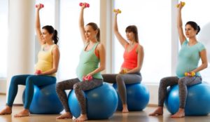
    All pregnant women, regardless of whether or not they lift weights, are at an increased risk for musculoskeletal injury. This is because the placenta produces a hormone called relaxin, which relaxes the body’s joints. This is critical since the pelvis needs to expand to accommodate the growing uterus and baby, and to allow the baby to pass through the birth canal during delivery.

    weight lifting is completely safe during pregnancy, as long as it is done in moderation. He also recommends sticking to weights under 30 pounds

    “Think bent over rows using a bench support and dumbells instead of a barbell and bending at the hips.”

    if you can hold a casual conversation during your workout, while feeling adequately challenged, you’re probably okay. Aim for breathing hard, but not feeling out of breath, and give yourself plenty of rest breaks for recovery during and between workouts.

    ABDOMINAL EXERCISES DURING PREGNANCY:-

    Myth: Stop training abs or you’ll risk separating your abdominal wall.
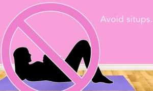
    Most article talk about stop abs completely, but as an experienced physio , I recommend doing certain abdomainal exercises for the following benefits:-

    1. Prevents excessive arching of the back, thereby reducing chances of back pain in post partun period.
    2. Improves core strength and reduces chances of Diastasis Recti
    3. More stability; during pregnancy our joints are more mobile making the body more flexible and prone to aches and pains, when u do weights, it just adds to the sense of stability and prevents these aches and pains.

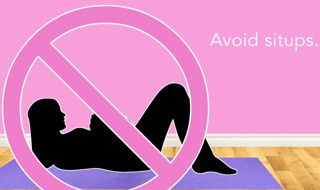
    Abdominal separation is a real thing that many women in pregnancy experience. It’s called diastasis recti (DR). DR occurs because of the growing fetus pressing onto your abdominal wall. Your rectus abdominals, the “6-pack” muscles, will stretch because of this, and the connective tissue holding them together can get very thin and soft.

    There are specific core exercises that you should not do that can increase the DR — for example, sit-ups, crunches, and front planks.

    What can you do? Half and tall kneeling positions, Pallof presses, dead bugs, farmer carries, diaphragmatic breathing exercises, and pelvic floor exercises.

    Remember to not breath hold during these exercises.

  • Pregnancy And Exercises

    Currently a very small % of women exercise at the recommended levels in India. In fact most obstetricians recommended avoiding exercise.

    There are lot of myths in terms of exercising during pregnancy, namely

    1. Physical activity has detrimental effects such as increasing body temperature.
    2. There is an increased risk of congenital abnormalities.
    3. Shifting oxygenated blood & energy away from the developing foetus, thus leading to disturbance in growth of the baby.

    All these and many more and no scientific research or proof to prove them true.

    Usually a normal pregnancy lasts for 36-40 weeks in which the woman gains around 10-15 kgs. The common physical problems associated with pregnancy are cramps, varicose veins, symphysis pubis dysfunction, carpal tunnel syndrome, rib pain, stress incontinence, tiredness & Low Back Pain

    Anatomical and biomechanical changes during pregnancy

    It is important to understand certain anatomical regions of the bones and muscles supporting the pregnant uterus as well their effects on our daily activities.

    The Pelvis

    The pelvic girdle is an important anatomical structure with respect to pregnancy. Relaxation of the pelvis is essential during pregnancy in order to make room for the growing fetus . The anatomy of the pelvic girdle as follows:

    • The pelvic ring includes the sacrum, the sacroiliac joints, the ilium, pubis and ischium and the pubic symphysis
    • Unlike the male pelvis which is narrow, deep and oblong the female pelvis is wide, shallow, roomy and round. The main differences are that the subpubic angle is wider and the inlet plane is bigger in the female pelvis.
    • In the pelvis the sacroiliac joints are the connection of the spine and legs
    • The sacro iliac joints (SIJ) are primarily stabilized by the anterior sacroiliac ligament, interosseous ligament and short and long posterior sacroiliac ligaments
    • Muscles that stabilize the SIJ joint are erector spinea, lumbar multifidi, abdominal muscle group (external and internal obliques, rectus abdominis and transverus abdominis) and hamstrings


    When the female pelvis does not conform to the normal alignment problems can occur. A non-optimal relation between the maternal pelvis and the fetal head may lead to complications during childbirth, potentially necessitating a caesarean section (Wilder 1988). Maternal causes include earlier pelvic trauma and innate malformations. Fetal causes include hydrocephalus disturbed circulation of cerebrospinal fluid which leads to brain dilation and cranial expansion.

    Pelvic floor muscles

    • The pelvic floor muscles are important during the pregnancy and the delivery.
    • The pelvic diaphragm consists of the levator ani and coccygeus muscles which support the pelvic viscera.
    • The coccygeus muscle in addition, flexes the coccyx. The pelvic wall consists of the piriformis and obturator internus muscles which also have a function at the hip joint.
    • The pelvic floor consists of the sphincter and erector muscles.

    Abdominal wall muscles

    • The anterior abdominal wall consists of the rectus abdominis muscle which acts to flex the trunk.
    • There are internal and external oblique muscles which act together to bend the trunk to the same side and rotate to the opposite side.
    • The obliques have a bilateral function to flex the trunk, compress abdomen and stabilize the pelvis.
    • These muscles are directed vertically, horizontally and oblique.
    • During pregnancy the diameter of the abdomen increases due to which some women develop a lateral separation of the rectus abdominus, which may persist even after the pregnancy.
    • The function of these muscles seems to be the same as with non-pregnant woman.
    • The ability of the abdominal muscles to stabilize the pelvis against resistance has been shown to be less in the third trimester of the pregnancy and this stays with the majority of the women until at least 8 weeks after birth.

    Biomechanics

    • During pregnancy the total body mass increases as the fetus develops, which increase the body weight and changes the physical loading.
    • As the gravid uterus increases in size, the centre of gravity shifts forward, causes the pelvis to move in the front and may result in an increased curvature in the lower back and anterior flexion in the neck .
    • Vertical forces applied to the feet during dynamic activities, such as walking, running and jumping, reach larger magnitudes ranging from two to five times body weight. However the majority of the mass is retained in the trunk region.
    • Getting up from a chair while pregnant causes a 33% increase in the tibiofemoral joint force, an 83% increase in patella femoral force, 100% increase in the activity of the quadriceps muscles and 35% increase in activity in the hamstring muscles.

    http://physiocure.in/updates/pregnancy-and-exercises/safety-issues-in-pregnancy/

    Effects of Exercises in Pregnancy


    [contact-form][contact-field label=”Name” type=”name” required=”1″][contact-field label=”Email” type=”email” required=”1″][contact-field label=”Message” type=”textarea”][/contact-form]

  • Tips To Prevent Neck Pain

    1. Avoid slouching during sitting, standing or lying down.
    2. Always try to keep your head in line with your shoulders and not let it rest forward.
    3. If you use an office computer, the desk should be at appropriate height such that the screen is at eye level. Also, the chair or the desk should have good forearm support available with shoulders relaxed at the side of the body.

    1. Sitting in front of the computer, try to sit with weight on the tail bone and not on your feet.
    2. If you use a pillow, it shouldn’t be too firm as the neck muscles are not relaxed. If it is too soft, the head sinks into the pillow and does not adequately support the neck during lying. The pillow should be such that it adequately provides support to the curve of your neckwhile sleeping.
    3. If you are used to sleeping on your stomach then avoid using a pillow as it bends the neck backwards causing pain. A pillow can be used when you sleep on your side.
    4. Take a break from using the mobile phone frequently. Change the hand while using the mobile. Using mobile stands which keep the neck at a comfortable angle rather than bent, helps avoid neck pain.
    5. While driving a car, the distance between the driving wheel and the car seat should be such that the elbows will be slightly bent.
    6. Use a pillow in the driver’s seat if you are not able to see the road while driving.
    7. If you carry a heavy one sided bag, change sides frequently as the neck and shoulder muscles of that side will become tight or may spasm leading to pain.

    8.If you carry a backpack, the strap height should be such that the backpack rests on your upper back         to avoid slouching.

    9.When you use a posture correction brace it helps with neck pain to some extent.

    [contact-form][contact-field label=”Name” type=”name” required=”true” /][contact-field label=”Email” type=”email” required=”true” /][contact-field label=”Website” type=”url” /][contact-field label=”Message” type=”textarea” /][/contact-form]

  • Tips To Prevent Back Pain

    Whether prevention is effective or not, depends on the person suffering from back pain and his/her motivation to deal with it actively. research for years has suggested that exercise along with education regarding lower back pain and ergonomic adjustments works just fine in solving the problems of back pain. so here are some tips to prevent the low back pain at all:-

    1. Sitting for long hours puts more pressure on the disc which may ultimately lead to back pain or disc prolapse. So it is important to change position every 15 mins to half an hour to reduce the load on the disc.

    2. If you are unable to move around and have to sit for a long time, doing buttock squeezes will activate the back and hip muscles.

    3. Use a foot stool and alternately shift weight if you have to stand for long hours.

    4. For housewives, when you cook in the kitchen, avoid leaning on the platform.

    5. Sitting in an office chair, use a lower back support rather than slouching.

    6. Frequently stand and bend backwards and forwards from your lower back and hips. This will help stretch out your back.

    7. Walk for at least 30 mins everyday.

    8. Reduction of fat around the abdomen helps reduce the strain on your lower back which helps reduce back pain.


    9. Proper weight lifting techniques. If you lift a heavy weight from the ground, let the object be in between your legs, squat and then lift it up.

    10. Pregnant women experiencing back pain can use a supportive belt which reduces the load of the pregnant uterus on the back.

    11. Take a strategic break in between your strenuous activities or matches in case you are a sports player.

    12. follow a Prehab for low back pain during your offseason period if you are a sports player


  • Shin Splints

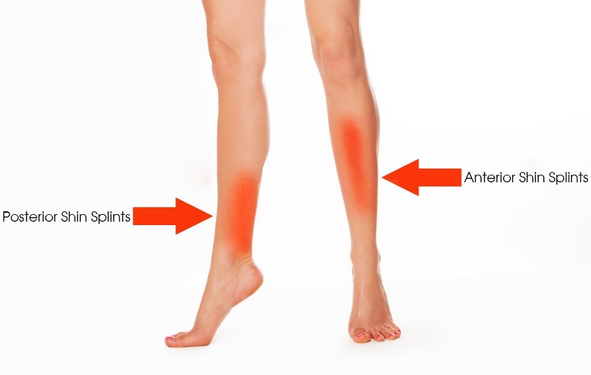
    Shin splints are usually caused more commonly in athletes. Clinically, it is also called a Medial Tibial Stress Syndrome’. Shin splints are pain that commonly occurs either in the front part or the inner part of the lower leg.                                                                   

    Shin splints pain causes are as following:-

    1. Flat foot
    2. Training errors including improper strengthening, pre-stretch or warm-ups
    3. Shoe design
    4. Type of road/surface
    5. Reduced bone mineral density
    6. Lack of appropriate recovery time

    The most common group of muscles implicated in shin splints are tibialis anterior, tibialis posterior, flexor digitorum longus, and soleus. These are the muscles that are present from below the knee and attach to the ankle joint and hence, also affect function at the ankle joint. Thus, if shin splints are not treated, they may cause long term changes in the biomechanics of your knee and ankle joint leading to other related problems.

    What are Shin Splints Symptoms:-

    Pain either in the front of your leg or the inner aspect of the calf. It is usually a dull aching pain that increases further with activity. Usually occurs during or immediately after the run and goes away if warm-ups are done. A lot of my clients ask

    Will shin splints cause swelling?

    My answer is yes and yes….first yes for mild swelling caused due to friction, second yes is for the occasional swelling in adjacent joints like in the ankle joint or the knee joint.

    Shin Splints Vs Stress Fracture:-

    If your symptoms are widespread and achy in nature, rather than a sharp pain, it could be a simple soft tissue injury. Generally, soft tissue injuries will respond well to the following:

     Ice therapy (15 mins a day, 2-3 times per day)

     Anti-inflammatories

     Stretching exercises

     Prescriptive loading exercises from a physiotherapist

     Offloading (no running)

    However, if you’re also experiencing the following, it could be a stress fracture, one of the most common worries for athletes:

     Pain at rest

     Significant increase in severity of pain with impact (weight-bearing > walking > running)

     Pain at night time

     Focal pain at a specific site along the bone

    then you must consult an orthopedic ASAP!!

    Red Flags:-

    Visit a physician immediately if you experience the below:

    1. If pain is at just one spot on the shin, it could be a stress fracture.
    2. If there is swelling, skin becomes shiny and there is excruciating pain, it could be leading up to compartment syndrome.

    Examination:-

    A proper detailed history of the conditions leading to the symptoms is to be known. For athletes, details about the training program are taken.

    The muscle strength and flexibility around the ankle and lower leg are to be evaluated. Your ankle, knee, and gait biomechanics are evaluated, and then accordingly, a treatment program is made.

    Treatment:-

    Initially, efforts are made to reduce the swelling and tenderness present. This can be done on Acute injury management principles using a combination of rest with cold therapy. Other electrotherapy modalities like TENS, iontophoresis, or ultrasound. Kinesiotaping is also used to provide rest to the muscles and for pain relief.

    Once the pain subsides, gentle stretching and strengthening exercises for the appropriate muscles are started. This is necessary to maintain the flexibility and to improve the biomechanics of the joint which was caused due to muscular dysfunction.

    here is one of the shin splints exercises to improve ankle mobility that we recommend for runners/athletes experiencing this injury. check it out!!

    Along with this, fascia and soft tissue release are also done to relieve any increased pressure within the leg and avoid further swelling. A proper strengthening program for the entire lower extremity including the core is essential and it helps in maintaining a proper posture and balance during running, jogging, etc.

    Later, progression is made to neuromuscular functional training which improves muscular dysfunction.

    Tips to prevent shin splints:

    1. Appropriate rest is to be taken before the next physical exertion.
    2. Regular foam rolling and/or self release using a tennis ball or a roller is a must for professional athletes.


    1. Increase ankle joint strength and flexibility through a training program
    2. Proper shoes are important and they should be reviewed every six months.
    3. It takes approximately 3-6 weeks to recover depending on the severity. So, early return to sports before this period may lead to development of chronic shin splints.