Blog

  • Tips To Deal With Post COVID Problems

    It’s almost a year since Coronavirus has taken the world for a 360-degree ride, spreading from one country to another, uprooting millions not only from good health but by wealth also. As an empath I really felt how can I help society, to help recover from this disease. definitely, I’m not the first line corona warrior, that would be your physician and nurses, but exercise is essential to get those lungs and other body parts functioning well.

    The most common symptoms of COVID-19 are

    • Fever
    • Dry cough
    • Fatigue

    Other symptoms that are less common and may affect some patients include:

    • Loss of taste or smell,
    • Nasal congestion,
    • Conjunctivitis (also known as red eyes)
    • Sore throat,
    • Headache,
    • Muscle or joint pain,
    • Different types of skin rash,
    • Nausea or vomiting,
    • Diarrhea,
    • Chills or dizziness.

    . Once stable and if indicated the main goal in respiratory physiotherapy is to mobilize secretions and ease the work of breathing

    Some people who have had COVID-19, whether they have needed hospitalization or not, continue to experience symptoms, including fatigue, respiratory and neurological symptoms.


    For all those individuals diagnosed with asymptomatic covid or mild covid problems, there are still some difficulties that may hinder you to spring back to normalcy once you have tested negative. they could be:-

    1. Fatigue
    2. Breathing difficulty in activities that require exertion
    3. weight loss causing general weakness
    4. mild aches and pains in different parts of the body
    5. stress/anxiety/depression.


    Research has quoted that

    Physiotherapy will have a strong role in providing exercise, mobilisation and rehabilitation interventions to survivors of critical illness myopathies associated with COVID-19 in order to enable a functional return to home.

    NOT RECOMMENDED IN EARLY STAGES:-

    In the early stages of COVID-19 and respiratory distress, care is advised when planning a treatment program. Common modalities often used by respiratory physiotherapists may be contraindicated in the acute phase as they may further compromise the increased work of breathing.

    Contraindicated interventions include:

    however one must take note that;

    Physio & yoga will have a role in the rehabilitation of COVID-19 patients who have not returned to their functional baseline, once they are no longer unwell

    So, to contribute to society, along with a dear friend and a skilled yoga practitioner Disha Gala, we have compiled a video to deal with few symptoms which may prevail post corona.

    In this blog and the video posted, we have presented a 5 part solution regarding the same as:

    1. Breathing exercises to improve Lung function
    2. Yoga poses to improve general mobility
    3. 6-minute walk test to determine the walking capacity
    4. Tips to deal with mental health issues
    5. Tips to ensure a nutrient-rich diet to recover from COVID symptoms

    watch this …..hope it helps

  • How To Deal With Ankle Sprains ?

    What is an ankle sprain?

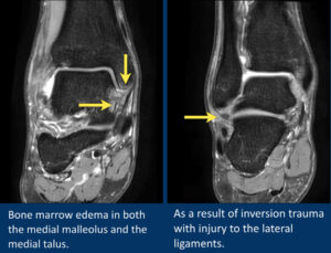
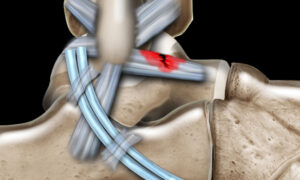
    An ankle sprain happens when your foot suddenly twists and stretches the strong bands (ligaments) that support your ankle beyond their limit. This usually affects the ligaments on the outer side of the ankle, causing pain, swelling, bruising and difficulty putting weight on the foot. Ankle sprains are extremely common in everyday life and in sports like marathon running, cricket, squash, tennis and dance.​

    How does an ankle sprain happen in daily life?

    In non‑athletes, ankle sprains often happen during simple day‑to‑day activities. Typical examples include missing a step on the staircase, slipping off the edge of a pavement, walking on uneven ground or stepping into a small pothole or gap. The foot rolls inwards, the ankle rolls outwards, and the ligaments on the outside of the ankle get overstretched or torn.​

    Sport‑specific mechanisms of injury

    In sport, the same basic twist happens, but often at higher speed and force:

    • Marathon runners: Fatigue, uneven roads, potholes and sudden changes in direction at aid stations can cause the foot to roll inwards during landing or push‑off, especially when the runner is tired late in the race.​

    • Cricketers: Bowlers and fielders are at risk when sprinting, cutting, turning quickly to chase the ball, or landing awkwardly from a jump or dive; landing on an uneven patch or another player’s foot is a common trigger.​

    • Squash, tennis and other racquet sports: Fast side‑steps, sudden stops, split‑steps and landing on an opponent’s or partner’s foot can all cause the ankle to roll sharply inwards. Video studies in court sports show many sprains happen when landing from a jump or changing direction with the foot fixed to the floor.​

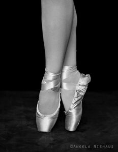
    • Dancers: Repeated jumps, turns and work on demi‑pointe/pointe put the ankle at risk when landing poorly, losing balance in turnout or rolling over the foot at the end of a jump

      In all of these, the common pattern is a sudden inward twist of the foot with the body weight moving over it too fast for the muscles to protect the ligaments.​

      What is a chronic ankle sprain?

      Sometimes the ankle never fully recovers from the first sprain, or it keeps getting sprained again and again. This is often called a “chronic ankle sprain” or “chronic ankle instability.” It means the ligaments and supporting muscles are no longer giving the joint the firm support and balance it needs, especially during quick or unpredictable movements.​

      Typical symptoms of chronic ankle problems include:

      • Repeated “giving way” or rolling of the ankle on uneven ground or during sport

      • A constant feeling of weakness or wobbliness in the ankle

      • Swelling or ache after walking, running, training or dancing

      • Stiffness or tightness, especially in the morning or after sitting

      • Fear or lack of confidence while running, jumping or changing direction

      Chronic ankle issues do not usually improve with rest alone. Without proper sports physiotherapy and ankle sprain rehab exercises, they can lead to more sprains, early joint wear‑and‑tear and ongoing pain.​

    Key sports physiotherapy principles :-


    After an ankle sprain, the goal is not to just “rest it” but to protect it briefly, control the swelling, and then start gentle, pain‑free movement as soon as it is safe. This early movement and gradual loading help the ligaments heal stronger and reduce stiffness.

    As the pain settles, the focus shifts to building strength, balance and coordination around the ankle, often using a brace or taping for extra support while you move and train. These exercises train the muscles and nerves to react quickly again, which lowers the chance of the ankle rolling over in the future.

    Instead of clearing you to play simply after a set number of days, your rehab should follow clear stages, from light everyday activity to full training and competition. At each stage your physiotherapist checks whether your strength, balance, hopping and sport‑specific skills match the other leg, and only then moves you to the next level or back to sport

    Sports‑specific biomechanics and pathomechanics

    • Marathon runners: Repetitive loading at mid‑stance and push‑off demands optimal dorsiflexion, elastic calf function and dynamic peroneal stability; limited dorsiflexion and poor neuromuscular control increase re‑injury risk, even when gross running gait looks “normal.”frontiersin+1
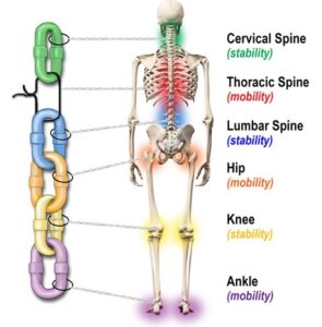
    • Cricketers: Bowlers and fielders experience high inversion moments during cutting, landing and boundary stops; lateral instability alters kinetic chain alignment, affecting knee, hip and lumbar mechanics during bowling and throwing.pmc.ncbi.nlm.nih+1
    • Squash/tennis/racquet sports: Multi‑directional lunges, split‑steps and rapid deceleration place high demand on frontal‑plane control; impaired proprioception and peroneal latency compromise change‑of‑direction mechanics and increase risk in side‑stepping and cross‑over steps.sciencedirect+1
    • Dancers: Repeated demi‑pointe, jumps and landings require end‑range plantarflexion control; lateral ligament injury disrupts alignment in turnout, landing mechanics and pointe control, increasing risk of chronic instability.pmc.ncbi.nlm.nih+1

    Acute vs chronic ankle sprain

    • Acute ankle sprain
      This is a “fresh” injury that has just happened, usually within the last few days or weeks.
      The main features are sudden pain, swelling, bruising and difficulty walking after you twist or roll your ankle. The focus here is ruling out a fracture, calming pain and swelling, and then starting early, guided rehab so it heals well.

    • Chronic ankle sprain / chronic ankle instability
      This refers to ankle problems that keep going for months, or repeated sprains over time.
      People often describe the ankle as weak, wobbly or “giving way,” especially on uneven ground or during sport. There may be on‑and‑off swelling or aching after activity, and confidence in the ankle is usually low. Chronic cases need a full strengthening and balance programme, sometimes extra bracing, and occasionally surgical opinion if rehab alone is not enough.

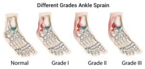
    Grading of acute ankle sprains (Grade I–III)

    Most research and clinical guidelines still use a three‑grade system to describe how badly the ligaments are hurt:

    • Grade I (mild)
      The ligaments are stretched but not torn.
      There is mild pain and swelling, but you can usually walk with only slight discomfort. Recovery is often quick with proper care.

    • Grade II (moderate)
      Some of the ligament fibres are torn.
      Pain, swelling and bruising are more obvious, walking is painful and the ankle may feel a bit unstable. Rehab takes longer and needs structured physiotherapy to fully restore strength and balance.

    • Grade III (severe)
      The ligament is completely torn, and sometimes more than one ligament is involved.
      There is significant pain, swelling and bruising, weight‑bearing is very difficult, and the ankle feels very unstable. Recovery is longer, and in some cases a specialist may discuss more intensive treatment or, rarely, surgery, along with a long, detailed rehab plan.

    Management of ankle sprains:-


    “Treatment is planned according to how new the sprain is and how badly the ligament is damaged, rather than using a ‘one size fits all’ approach.”


    Type / Grade What it means (simple) Main goals of treatment Typical support & activity level
    Acute – Grade I (mild) Ligament is stretched but not torn; mild pain and swelling; you can usually walk with some discomfort. Calm pain and swelling quickly, protect the ankle briefly, then start gentle movement and gradual loading so it does not get stiff or weak. Short period of protection (brace/taping), home care for swelling, early comfortable movement, then gradual return to walking, daily tasks and light sport.
    Acute – Grade II (moderate) Some fibres of the ligament are torn; more pain, swelling and bruising; walking is painful and ankle may feel a bit unstable. Protect the joint while it settles, then carefully restore full movement, strength and balance so the ankle feels stable again. Stronger external support (brace/taping, sometimes crutches for a few days), clinic‑based rehab, step‑by‑step return from daily activities to jogging and then sport, usually over several weeks.
    Acute – Grade III (severe) Ligament is completely torn (often more than one); marked swelling and bruising; very hard to put weight on the foot. Give the ligament time to heal in a protected position, then follow a long, structured rehabilitation plan to regain movement, strength, balance and confidence. Firm support (boot or rigid brace) and reduced weight‑bearing at first, followed by closely supervised rehab; gradual return to normal walking, then sports; sometimes a specialist opinion if the ankle stays very unstable.
    Chronic ankle sprain / chronic instability Ankle feels weak or “keeps rolling” months after the first injury, or there are repeated sprains. Improve long‑term stability, reduce “giving way,” and prevent further sprains by training the muscles, balance and control around the ankle. Detailed assessment, longer rehab programme focusing on stability and control, possible regular use of brace/taping for higher‑risk activities, and ongoing “maintenance” exercises to keep the ankle strong.

    Sport‑specific rehab and return‑to‑play

    Sport Rehabilitation Focus Return-to-Play Principles
    Marathon Runners Early gradual return to running, progressive walking to jogging, strength building for calf muscles, improved balance and coordination to support running mechanics even if gait looks normal Gradual increase in running load and pace, ensuring pain‑free movement and stability in running pattern before full return
    Cricketers Phased running drills starting with straight-line running progressing to curves, cutting, sliding, and landing; strengthening of trunk and hips to support complex movements like bowling and fielding Systematic progression through cricket-specific movement patterns, working towards full bowling and fielding actions under supervision
    Racquet Sports (Squash, Tennis, etc.) Focus on single-leg strength, lateral balance and agility, multi-directional movement drills including split steps and lunges tailored to court demands Increasing intensity of court-specific drills and match-like movements only after achieving adequate strength, balance, and control
    Dancers Gradual progression from controlled barre work to centre stage, enhancing single-leg endurance, landing control, and supporting complex turns and jumps Slowly advancing to full choreography and performance, often with external support (like taping) during early return phase to reduce risk of recurrence
    All Sports Supervised physiotherapy focusing on neuromuscular training and proprioception (body awareness), with clear, objective criteria to measure progress before returning to sport Phased rehabilitation models emphasizing return to activity, sport, play, and competition steps using strength, balance, hop, and sport-specific functional tests

    References:-

    1. https://pmc.ncbi.nlm.nih.gov/articles/PMC164373/
    2. https://www.massgeneral.org/assets/mgh/pdf/orthopaedics/sports-medicine/physical-therapy/rehabilitation-protocol-for-ankle-sprain.pdf
    3. https://www.sanfordhealth.org/-/media/org/files/medical-professionals/resources-and-education/014000-01095-flyer-ankle-sprain-rehabilitation-pt-guideline.pdf
    4. https://patialaheart.com/blog/ankle-sprains-diagnosis-treatment-and-rehabilitation-for-athletes/
    5. https://www.physio-pedia.com/Ankle_Sprain
    6. https://ijspt.org/wp-content/uploads/2024/06/8-Flore.pdf
    7. https://www.massgeneral.org/assets/mgh/pdf/orthopaedics/foot-ankle/pt-guidelines-for-ankle-sprain.pdf
    8. https://www.narayanahealth.org/blog/rehabilitation-for-ankle-sprains-everything-you-need-to-know
    9. https://www.orthobullets.com/foot-and-ankle/7028/ankle-sprain
    10. https://orthoinfo.aaos.org/en/recovery/foot-and-ankle-conditioning-program/
    11. https://www.aafp.org/pubs/afp/issues/2001/0101/p93.html
    12. https://emedicine.medscape.com/article/1907229-treatment
    13. https://www.orthobullets.com/foot-and-ankle/7028/ankle-sprain
    14. https://www.nata.org/sites/default/files/2025-08/ankle-sprains.pdf
    15. https://www.europeanreview.org/wp/wp-content/uploads/1876-1884.pdf
    16. https://www.frontiersin.org/journals/medicine/articles/10.3389/fmed.2022.868474/full
  • Knee Rehabilitation From Physio’s Perspective – Beginner’s Level

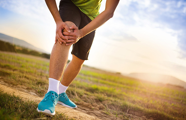
    Anyone can be a victim of a knee injury; whether you are a professional athlete, a weekend fitness buff or someone just busy carrying out your day-to-day activities. The knee joint is unique in that it carries body weight at the same time as it provides flexibility and mobility. It is formed by the femur, the tibia and the patella and held together by muscles, ligaments and tendons. This construction predisposes the joint to risk of injury from sudden movement and repeated wear and tear.

    Some common knee injuries

    • Ligament sprains– ligaments are bands of strong tissue that stabilize the knee joint together. Overstretching can cause the ligament fibre to tear and bleed into the surrounding tissues, causing pain, swelling and a feeling of ‘giving way’. Severe tears like ACL Injury may require surgery.
    • Cartilage Tears – cartilage also helps stabilize and protect the knee joint. Pressure from twisting and turning during weight-bearing exercise like squash may tear the cartilage, causing pain, swelling and locking of the knee joint leading to problems like Chondramalacia patellae
    • Tendon Strains – tendons are cord-like bands that connect muscle to bone. The patellar tendonconnects the kneecap (patella) to the tibia (shin). Patellar tendonitis or ‘jumper’s knee’ is inflammationof the patellar tendon. This is an overuse injury commonly caused by repeated jumping, as in
    • Patello-Femoral Syndrome – excessive friction between the surface of the patello-femoral joint (kneecap) and the femur (thigh) can result in knee pain. It may have a number of causes and should be assessed by your physiotherapist.

    Mechanism of injury:-

    Knee injuries can be caused by trauma, such as an accident or a fall, or even by a sudden increase in your daily activities. Other injuries develop over time, from factors including structural problems within the joint itself, incorrect athletic training methods or technique, and poor equipment.

    • Footwear –a new pair of athletic shoes or a change to heels instead of flats can affect the way you walk and, in turn, can place stress on the muscles and tendons around the knee joint. selecting The Running Shoe should be carefully thought about
    • Frequency and intensity of a regular activity – Any change such as jogging or playing tennis on a different surface can affect the knee joint. For example, simply moving the activity from a soft track or clay court to a hard surface such as a road or hard court can add stress to the knee joint and increases the risk of injury.
    • Compensation for an injury elsewhere. A blister on your foot, for example, may cause you to change the way you step to minimize the pain, thus placing a strain on the knee.

    MANAGEMENT OF KNEE PAIN:-

    how effective is physio in problems like knee osteoarthritis?

    There is good evidence to support the physiotherapy management of knee OA. The main areas supported by research include:

    Physiotherapy vs arthroscopy for knee OAphysiotherapy exercise and education, combined with standard medical care (simple pain medication) is equally effective to keyhole knee surgery (arthroscopy) in the management of knee OA. In people with moderate to severe knee OA, it has been found that arthroscopy added no additional benefit in terms of physical function, pain or quality of life, when compared to physiotherapy and simple medication. In addition, physiotherapy has been shown to have minimal risk of negative side effects compared to surgery.

    How to Prevent Knee Injuries

    Obviously it’s impossible to completely eliminate knee injuries, but athletes can take proactive steps to keep their knees healthy and strong. Follow these three guidelines in your training program:

    • Develop lower body strength for additional joint stability. The hamstrings are a particular cause for concern, because weakness there is associated with a higher probability of ACL injuries. Perform exercises like Squats, Deadlifts and RDLs, which develop the quads, hamstrings and other lower body muscles for balanced muscle strength.
    • Develop proper technique for running, jumping and other lower body skills
    • Perform plyometric exercises to improve technique and muscle power, teaching muscles to fire and stabilize the joint immediately after the foot touches the ground

    Treating knee Injuries:-

    Treatments are usually injury specific. In acute stages ice, rest from activity, control excessive swelling and unload the joint with crutches or cane. Bracing or splinting can be useful if there is instability.

    We recommend to avoid anti-inflammatories, as they may interfere with the necessary stages of healing, which includes a healthy inflammatory response. Early range of motion activities and other light activities are encouraged.

    At Physiocure we work at resolving these knee injuries by following a structured rehab programme which will include the following goals:-

    • Reduce pain
    • Restore Mobility
    • Develop control and strength
    • Improve and maintain alignment
    • Return to sport/work

    When Should You Consult a Physiotherapist?

    If you some exercises/workout and they increase your knee pain or the condition doesn’t improve, a physiotherapist can help design a more personalized exercise prescription for you.

    It is also important to note that there are many different conditions that can produce knee pain. Therefore, one must be careful to self-diagnose with any particular knee injury and seek professional help to get the correct evaluation and appropriate treatment.

    As a general rule, I recommend seeking treatment for any injuries that have not resolved within two weeks.

    https://www.youtube.com/watch?v=uwfPJMxf6Rc

    We cater to knee injuries like:- ACL InjuryMeniscal InjuriesChondramalacia patellae, Patellofemoral Pain SyndromePatellar tendinopathy

  • Sports Physiotherapy: Why Modern Athletes Are Choosing Evidence-Based Rehab

    If you play a sport — whether it’s Cricket at Juhu Beach or Balkanji bari turf or at MIG Cricket Club in Bandra East, Badminton at Willingdon Gymkhana or in Prabodhankar Thackeray Krida Sankul for Pickle ball, football turf sessions at TurfPark | St Andrews, or Early-morning Marathon training at Carter Road or with STriders or mUmbai runners— chances are your body is dealing with far more load, impact and repetition than you realise.

    And with increasing sports participation in Mumbai, sports injuries are becoming extremely common:
    knee pain while running, shoulder pain while smashing a shuttle, ankle sprains while playing football, lower-back stiffness from gym training.

    This is exactly where sports physiotherapy makes the difference.


    Why Sports Physiotherapy Is Not the Same as General Physiotherapy

    A lot of people still believe physiotherapy is only for pain relief.
    But sports physiotherapy works differently — it focuses on movement, muscle coordination, sport-specific loading, and performance.

    Unlike general physiotherapy, sports physio looks at:

    • What movement pattern caused the injury

    • How your biomechanics affect performance

    • What strength or mobility deficits created overload

    • Whether you are loading too much, too soon

    • How to return you to sport safely

    Most of the times I have noticed atheletes or weekend warriors playing with injuries. they just dont feel like/ dont want o/ dont have time to get it assesed. many a times they visit the medical doctors take basic meds and dont adress it completely and then once the tournament or pain has subsided they dorectly hit the gym and expect gym trainers to do the therapy they should have gone to in the first place.

    A sports physiotherapist bridges that gap by keeping training:

    ✅ Safer
    ✅ Smarter
    ✅ Stronger
    ✅ More sustainable


    Most Common Sports Injuries Seen in Mumbai Athletes (2025 Trends)

    Across clinics in Santacruz, Juhu, Bandra and Andheri, the most common injuries reported by athletes today include:

    1️⃣ Knee pain in runners

    • IT Band syndrome

    • Runner’s knee

    • Patellar tendinopathy

    2️⃣ Shoulder injuries in badminton & tennis players

    • Rotator cuff irritation

    • Shoulder impingement

    • Scapular muscle weakness

    3️⃣ Ankle sprains in footballers & recreational athletes

    • Lateral ligament sprains

    • Balance/proprioception deficits

    4️⃣ Tendon injuries in gym-goers

    • Biceps tendinopathy

    • Achilles overload

    • Elbow tendon pain

    5️⃣ Lower-back tension due to poor mobility

    Especially in cricketers (fast bowlers) and people doing heavy lifts.

    The trend across all areas?
    Most injuries are preventable with early load management, movement correction and sport-specific rehab.


    What a Sports Physiotherapist Actually Does (Beyond Pain Relief)

    A modern sports physio today uses an integrated approach:

    https://www.instagram.com/physiocure_sportsrehabclinic/reel/DGnJ-BAM54P/

    ✔ Movement Screening

    To analyse running form, jumping mechanics, landing technique, racket swing pattern, treadmill gait, etc.

    ✔ Manual Therapy

    Soft-tissue release, joint mobilizations, and myofascial techniques to restore mobility.

    ✔ Strength & Conditioning

    Structured progression for:

    • Strength

    • Mobility

    • Power

    • Agility

    • Endurance

    starting 2025 we conduct only strengthe and conditioning classes at Physiocure. we conduct Mat Pilates on monday, wednesday and fridays and we conduct align and activate posture fitness class on tuesdays and thursdays. you can call us for further details.

    ✔ Sport-Specific Rehab

    Every sport in Santacruz/Bandra/Juhu has unique demands:

    Cricket: rotational core strength, shoulder integrity
    Badminton/Tennis: upper-body control, footwork mechanics
    Running: lower-limb load tolerance, hip stability
    Football: agility, ankle stability, hamstring strength

    ✔ Return-to-Sport Testing

    A critical but often ignored step that prevents re-injury.

    Hop tests, gait analysis, Y-balance tests, sprint tests — depending on the sport.


    Why Athletes from Santacruz, Bandra & Juhu Actively Seek  Affordable Sports Physiotherapy Today

    1. Turf culture

    Football turfs across Santacruz & Bandra have exploded in popularity, increasing high-speed injuries. we can help turf footballers continue playing while we help them recover through sports injuries smoothly

    2. Running communities

    Running clubs like Striders, Bombay running, Nike running club, Mumabi Road Runners, Asics Runners club and self practicing runners around bandra, Khar, santacruz dont know that we run a runner specific injury prevention prorgram. we also do Normatech leg recovery sessions for post marathon or on regular trainig days and help with all kinds of running injuries.

    3. Fitness studio boom

    witht he changing trends some really cool multifunction gyms like FLOFIT BOX  in Bandra and MMA Matrix centre in Khar/ sanatcruz linking road and some really cool gyms around sanatcruz like Fitness First, dessus fitness , Transform gyms and some yoga centres like the body temple yoga, we definetly know people here like to go to the fitness centres and many a time they may need a sports physio for them to address the aches and niggles. we are happy to serve all kinds of fitness freaks so they continue their fitness journery pain free.

    4. Competitive amateur athletes

    Badminton players, tennis enthusiasts and cyclists train hard but recover poorly — leading to breakdown.

    5. Early rehab awareness

    People now realise physio is not last-minute treatment — it’s performance enhancement.


    2025 Insights: What The Latest Research Says

    Sports physiotherapy worldwide is shifting to:

    📌 Load-based tendon rehab (not rest)

    Achilles, patellar & shoulder tendons heal through progressive loading — not through avoiding activity.

    📌 Strong evidence for isometrics + eccentrics

    Particularly in runners and racket-sport players.

    📌 Mobility + control > flexibility alone

    High-performing athletes don’t just stretch — they stabilise.

    📌 Strength symmetry before return-to-sport

    Especially after ACL and ankle injuries.

    📌 Train the movement, not the muscle

    Sport-specific rehab gives faster results than isolated strengthening.


    Sports We Commonly Work With (Based on Real Clinic Cases & Field Trends)

    From everyday athletes to competitive players, sports physio in Santacruz commonly manages:

    🏸 Badminton

    Shoulder overload, knee pain, plantar pain, elbow strain.

    🏃 Running / Marathon Prep

    ITB pain, Achilles tendinopathy, shin splints, calf tightness.

    🏏 Cricket

    Lower-back stress, shoulder issues, hamstring pulls.

    Football

    Ankle sprains, groin strain, hamstring imbalance.

    🏋 Gym / Strength Training

    Lifting injuries, mobility deficits, core weakness.

    🎾 Tennis

    Elbow tendon pain, shoulder dysfunctions, wrist loading issues.


    How to Choose the Right Sports Physiotherapist in Santacruz / Juhu / Bandra

    Before starting rehab, ensure your physio:

    ✔ Understands sport-specific biomechanics
    ✔ Offers structured progression plans
    ✔ Uses objective testing (strength & functional)
    ✔ Treats the root cause, not just the symptoms
    ✔ Blends manual therapy + strengthening + movement retraining

    A good sports physio is not someone who gives only massage or only exercises —
    but someone who can combine science, coaching and clinical expertise.


    Final Thoughts

    Sports physiotherapy is no longer limited to elite athletes — it is essential for anyone active in today’s high-demand fitness culture.

    Whether you’re a:

    • Runner at Juhu Beach

    • Cricketer in Santacruz

    • Badminton player in Bandra

    • Footballer at Carter Road

    • Gym enthusiast training in Khar

    …your body deserves the same level of care that professional athletes receive.

    Your performance, recovery, and longevity in sport depend on how well you move — and how smartly you train.
  • What Examination To Expect In A Physio Clinic ?

    This is a very common question that haunts a lot of people who visit a physio clinic for the first time or are visiting us for the first time. As an experienced therapist, i believe, a proper examination will , not just on the first visit but on each and every visit , helps us identify the problem areas , help chart out the course of treatment and  inturn help design the best possible rehab plan for your existing issues.

    Why does a physio need to examine, when the ortho has already given the diagnosis?

    this is a very common question i’ve heard from only couple of my clients who got frank about their view about physiotherapy practices. so here is my explanation. when a physio examines a client with a neck pain or low back pain or knee pain, we are looking at structures involved in causing the dysharmony in the movements and how they can be corrected.


    This is why clinical expertise in assessing the problem is important. through experience ,the physio can identify the structure contributing to the pain, problem and design a treatment plan.

    On the first visit, we will analyse your history and current situation by understanding

    • Where you are having the problem
    • How did the problem start
    • How the problem is affecting your ability to do daily activities
    • Your past medical history and any medications you are taking
    • A detailed physical examination
    • Checking your posture
    • Examination of range of movement
    • Testing of movements specific to your problems & muscles strength

    Functional tests specific to your problem like balance, walking patterns or how you carry load

    Sometimes the assessment may be completed in 2- 3 sessions, depending on the severity of the pain or disability.

    Once the assessment is complete, a diagnosis is made and we usually explain the problem to the client and how it will be dealt with from our side.

    Quote

    “Either you can do guesswork, or examine the problem….we choose the later

    if you want to visit us fillout this form and mail it on info@physiocure.in

    General Assessment Form

    follow us on insta to get regular updates about our work and understand how we carry assessments in certain clients.

    https://www.instagram.com/p/B9qZhcNAJmn/

  • Post Natal Back Pain – A Personal Take

    Its been a while since I wanted to share my personal experience of dealing with post pregnancy back pain and finally I decided to share my experience .Post Natal Back pain has been a trouble to 90% of the moms and its been their reason of being afraid to exercises too soon or take up fitness like before their pre preganacy days.


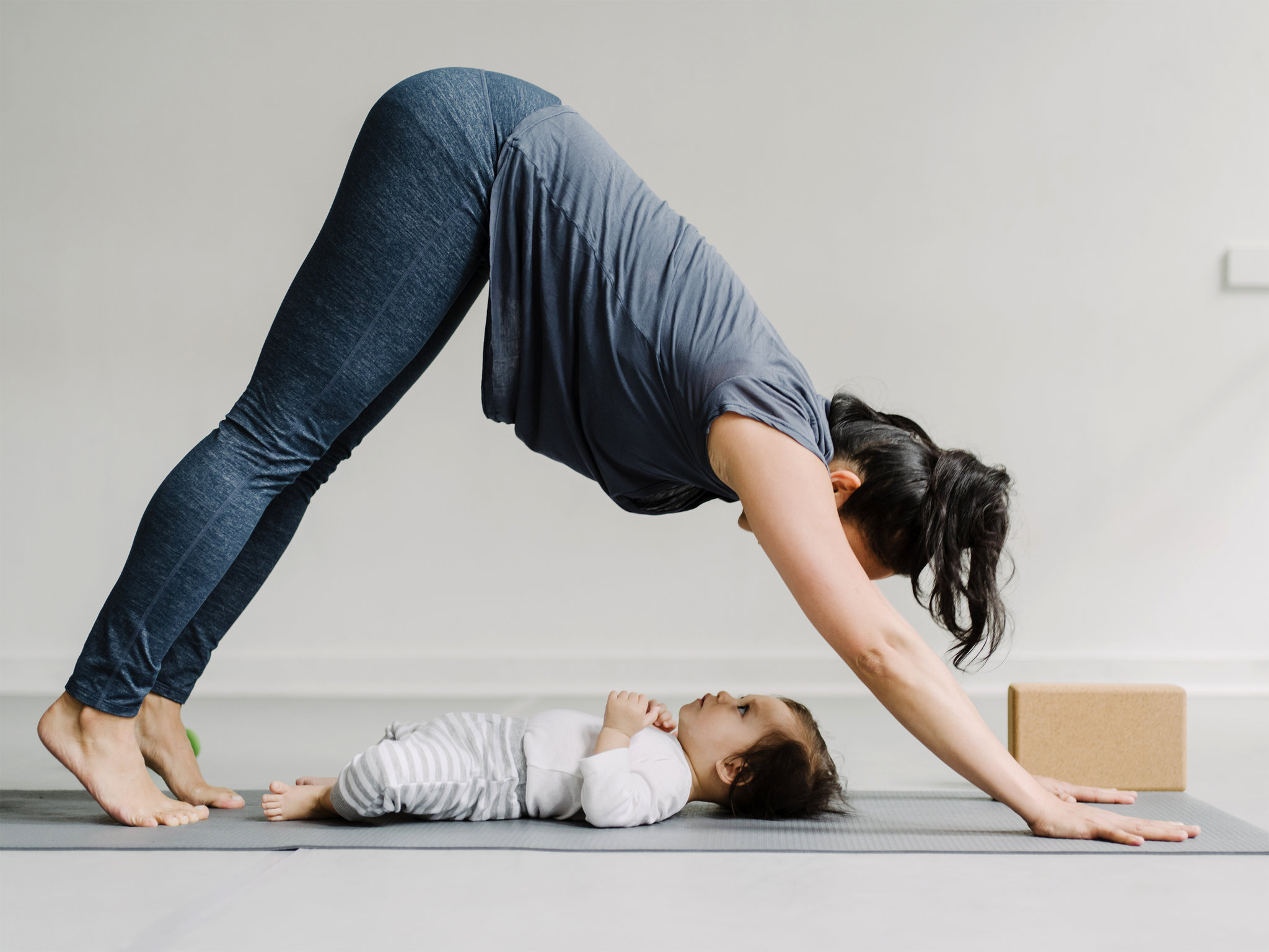
    postnatal fitness


    Its a beautiful feeling to have your baby finally in your arms but , its a difficult time immediately post pregnancy. there is no day and night schedule for the first 40 days ( like they say in traditional Indian words to which I totally agree), you need to recover out of the birthing experience yourself, you need to get to know your baby more (no doubt your partner and family support is always there) but still its a different experience.

    For me, it was a C-section or a planned delivery and I started experiencing back pain second day post delivery. it was really difficult to turn sides, get up from the lying down position. if I got up from the chair, I would be bent for a while and then gradually straighten up. once I lied down after feeding my baby in sitting position, even lying down flat would be uncomfortable. somewhere it struck me that the earlier the measures are taken to solve the back pain, the better will be the recovery.

    I started my back care after getting discharged from the hospital, at home basically. I started hot compress initially, atleast 3-4 times a day. there where medications to deal with the pain for the first 10 days as well. I did not follow any exercises for the first 2 days in the hospital.

    As soon as my baby delivered on the 3 rd day , I started with this simple exercise to activate lower back muscles along with pelvic floor muscle training along with hot compress and started to feed my baby in sitting position most of the times.

    Its very important to take a good back support while feeding the baby and avoid slouching as it aggravates the lower back pain.

    At this point every time, I sat for nursing , I would ensure there is sufficient back support and i’m not sitting uncomfortably in a wrong posture to feed. it the most important thing to follow till u continue feeding your baby.

    At 2 weeks, I started with these lower back = beginner level rehab exercises. These worked fantastically to activate my lower back muscles. I would do the following exercises everyday in the morning post a traditional Indian massage along with PFM’S each for 10 repetitions daily for the next 2 weeks.

    At 4 weeks, I started with Mckenzie back extensions, supine bridges, level 0 and Level 1 core muscle exercise along with 20 min leisure walks daily to add on the previous program. I continued taking hot compress at least twice in a day.

    As a traditional Indian measure, I had also started massage with a local masseur, which helped relax the back every day. I also had a few diet changes to support my recovery which were majorly traditional Indian measures.

    Normally most informative websites will advice to start exercising only after 8 weeks of pregnancy. but the latest research says otherwise, the earlier you start doing easy and low key exercises the faster you will recover from back pain due to pregnancy

    At 8 weeks, I was doing half an hour walks daily along withe these core strengthening for lower back  exercises.

    Currently I occasionally experience back pain, to be frank, sometimes it is not possible to keep the schedule as i’m still learning the art of multitasking with baby, work and house responsibilities, all in one hand.

    I think what really helped me is all the activity during my pre natal time. being active, to not stop working or not to be adviced bed rest, to do squats during pregnancy were few things helped me get back to fitness faster.

    currently i’m following this core stability program which is based on Pilates school of thought to keep my lower back strong. Check it out !!

    If you have faced a similar problem do contact me at physiocure17@gmail.com, I maybe able help you out.

    PS:- share this article and follow the blog post for further updates on regaining my fitness post pregnancy.


  • Shoulder Pain

    Shoulder pain is a common problem which lot of regular or athletic population face during their life time. However, identifying the source of this shoulder pain can get better insights to the resolution of the problem.

    shoulder pain
    shoulder pain

    Understanding shoulder pain

    This requires knowledge of the structures involved the activity which lead to the pain and the present or past history of events.

    For example, a regular individual who complains of shoulder pain may have Sub acromial impingement syndrome or rotator cuff syndrome or a cervical disc prolapse, it’s the occupation, lifestyle presenting posture of the individuals, and many other factors point out to the cause of pain and the provisional diagnosis.. At the same time it is important to judge and understand that the shoulder pain could be referred from the heart ache as well.

    SHOULDER INJURIES IN SPORTS:-

    • The shoulder is a frequent site of injury in competitive athletes. Shoulder injuries
      comprise between 8% and 13% of all athletic injuries.
    • These injuries may result from repetitive overhead activities (in swimmers, baseball pitchers, tennis players, and javelin throwers) or direct trauma (in football and rugby players).
    • Within a given sport, most shoulder injuries are position specific. In baseball, shoulder injuries are more common among pitchers. In swimming, shoulder injuries are most common in those who compete in butterfly, freestyle, and backstroke events.
    • In addition to the nature of the sport and the position played, shoulder injuries in the athlete depend on the anatomic variants specific to each athlete, their age, conditioning, level of play, level of overhead activity, techniques used, and the length of participation in their respective sport.

    THE SHOULDER COMPLEX AND ITS FUNCTION:-

    • The shoulder Complex is made up of these 4 joints which are run by over 30 different muscles. the sternoclavicular (SC),acromioclavicular (AC), and glenohumeral (GH) joints, as well as the scapulothoracic articulation..
    • Over a period of years, there certain wear and tears occuring around these joints which disrupt the normal working harmony of these joints and the muscles
    • The primary goal of the shoulder complex is to position the hand in space for activities of daily living.
    • Secondarily, during overhead athletic activities such as throwing and serving, the shoulder functions as the “funnel” through which the forces from the larger, stronger muscles of the legs and trunk are passed to the muscles of the arm, forearm, and hand, which have finer motor skills.
    • The ability to execute these actions successfully comes from a balance in mobility and functional stability of the shoulder complex.
    • The majority of the stability at the shoulder is determined by the soft tissue structures that cross it. The ligaments and capsule form the static stabilizers and function to limit translation and rotation of the humeral head on the socket of the shoulder joint.
    • The muscles make up the dynamic stabilizers of the shoulder joint and impart stability in a variety of ways. During muscle contraction, they provide increased capsule ligamentous stiffness, which increases joint stability.
    • They act as dynamic ligaments when their passive elements are put on stretch . Most importantly, they make up the components of force couples that control the position of the arm and the shoulder blade, helping to appropriately direct the forces crossing the shoulder joint .
    • Proper scapula (shoulder blade) motion and stability are critical for normal shoulder function. The scapula forms a stable base from which all shoulder motion occurs, and correct positioning is necessary for efficient and powerful arm movement.
    • Abnormal scapula alignment and movement, or scapula thoracic dyskinesis, can result in clinical findings consistent with instability and/or impingement syndrome.

    WHAT DO SYMPTOMS POINT OUT TO?

    • Weakness may appear as a true loss of power, suggesSai RCfting a neurological disorder, or as a sudden and surprising inability to abduct the shoulder – perhap sdue to a tendon rupture. Between these extremes there is weakness in performing only certain movements and weakness associated with pain.
    • Instability symptoms may be gross and alarming(‘my shoulder jumps out of its socket when I raise my
      arm’); more often they are quite subtle: a click or jerk when the arm is held overhead, or the ‘dead arm’ sensation that overtakes the tennis player as he or she prepares to serve.
    • Stiffness may be progressive and severe – so much so as to merit the term ‘frozen shoulder’.
    • Swelling may be in the joint, the muscle or the bone; as a patient its difficult to know the difference.
    • Deformity may consist of asymmetry of the arm, squaring of shoulders, winging of the
      scapula or an abnormal position of the arm.
    • Loss of function is usually expressed as difficulty with dressing and grooming, or inability to lift objects or
      work with the arm above shoulder height.


    These are the most common signs and symptoms and each points towards involvement of specific structures in the shoulder which help aid our diagnosis.

    WHAT SHOULD U EXPECT DURING YOU EXAMINATION?

    • once you contact us, we might have a brief chat about your problem on the phone.
    • on the first visit, you should get all your investigations as well as referal papers.
    • on the first visit maximum time goes in understanding how did the pain start and a brief history is taken, all the reports are read and an examination pertaining to your shoulder pain is performed.
    • then i usually explain all my clients the course of treatment, all of this will take around 20 min.
    • next the treatment is started on the first visit itself.

    WHAT TREATMENT PRINCIPLES BASED ON DURING SHOULDER PHYSIO?

      • In most patients, rehabilitation after a shoulder injury should initially focus on pain control and regaining the coordinated motion throughout all components of the shoulder complex.
      • Once motion is regained, attention is shifted to strengthening and re-educating the muscles around the shoulder to perform their normal tasks.

    • To reproduce the precision with which the shoulder complex functions, the muscles need to be re-educated through “learned motor patterns.”
    • These patterns position the shoulder complex in “predetermined” ways and activate the muscles in precise synchronization to maximize recovery of function.
    • Associated conditioning of the lower extremities and trunk muscles is extremely important because over 50% of the kinetic energy during throwing and serving is generated from the legs and trunk muscles.
    • Therefore, rehabilitation of all components of the kinetic chain is required before the successful return of competitive or strenuous overhead athletic activities.
  • Rotator Cuff Injuries

    With advance in newer treatment techniques, it is imperative as a physio for me to make you understand different kinds of shoulder pains. Another reason to pursue this topic is to provide readers help for rotator cuff pain and understand the differences in rotator cuff pain vs frozen shoulders. In this article we will be discussing mainly about Rotator cuff injuries (RCI)  which occur in normal individuals. RCI includes the terms tendinitis, tendinosis, tendinopathy, partial thickness tear(PTT), full thickness tear(FTT) used for the rotator cuff injuries.

    There is an increased incidence of shoulder pain in individuals who participate in repetitive overhead activities including repetitive throwing sports like baseball or volleyball, occupations such as painting or carpentry. Rotator Cuff Tendinopathy(RCT)  is seen as the most common type of shoulder pain as it is seen in about 30% of the overall population.

    RCT incidence also increases with age, which needs to be considered when we talk about Rotator Cuff Injuries.

    Before getting to the details of this syndrome, its necessary to know about the anatomical structures involved.

    ANATOMY AND BIOMECHANICS OF ROTATOR CUFF:-

    In earlier days the area of pathology was identified mainly affecting the subacromial arch, the sub acromial bursa, the supraspinatus tendon. Recent research, adds to the affection of rotator cuff also. These rotator cuff muscles get pinched between the bones that form your shoulder leading to shoulder pain, this is a basic defination of RCS

    The Rotator Cuff (RC)

    The RC comprises of 4 muscles namely; the supraspinatus, subscapularis, infraspinatus and teres minor tendons. These RC tendons blend together with the biceps, the coracohumeral ligament and the capsule (Matava, Purcell & Rudzki, 2005 p. 1405-6). They form an aponeurotic tendon surrounding the head of the humerus that contribute to the stability, movement and sensory motor control of the GHJ.

    This structure helps the RC to resist failure, since load is distributed over a wider area.

    Rotator cuff tendon structure:-

    rotator cuff pain location

    Also known as the rotator cable described as a point of attachment of 4 rotator cuff muscles onto the arm bone. (The white area where the muscles are ending in the picture.) This area is a critical zone as the blood supply is limited and hence when subject to wear and tear it tends to develop tear and contributed to rotator cuff pathology.

    CAUSES OF RCS:-

      • Intratendinous degeneration or tendinosis might occur as a result of the combination of limited vascularity in Codman’s critical zone and external compression of the tendon structures in the subacromial space’
      • As these tendons become weak, indirectly the function of the rotator cuff muscle gropu becomes inefficient/ weak which affects the joint normal biomechanics. What happens is everytime the arm is lifted, the arm bone (humerus) tends to ride up hitting the sub acromial bursa/acromion bone, causing irritation.
      • Nho et al. (2008, p. 988) suggest the influence of age and activity on the shape of the acromion, as well as environmental causes such as age, co-morbidities (like diabetes mellitus and smoking) and overuse in impairing the healing processes of the tendon.
      • It seems that superior translation of the humeral head plays a significant role in the pathology (Lewis 2009a, p. 239).
    • Chronic impingement can lead to bursal inflammation, tendonitis and can eventually cause degenerative changes and tears of the cuff tendons if not treated (Lin, Weintraub & Aragaki 2008, p. 627; Nho et al. 2008, p. 988).

    In summary  the etiology of Rotator Cuff disease is multifactorial, including both intrinsic and extrinsic factors. It is believed that articular side tears are associated with intrinsic changes to the tendon and bursal side tears associated with SA impingement

    Signs & Symptoms:-

    RC pathologies (including SAIS) can manifest in anterolateral shoulder pain

    • patient usually grabs the whole shoulder when describing the location
    • aggravated in overhead activities,
    • described as constant ache and
    • night pain.
    • Pain may radiate to the scapula/ neck/ elbow/ fingers, not in a dermatomal way

    Examination

    A thorough physiotherapy objective examination includes few rotator cuff syndrome tests:-

    • Testing of the neck including ROM, palpation and provocation tests are done to rule out referred pain.
    • The shoulder girdle is examined for atrophy, weakness and for shoulder blade and arm muscles asymmetry
    • Active and passive shoulder ROM is assessed (where in we find that limited Active should er movements but passive movements are full
    • strength in all planes is tested once the pain is in control.


    • Tests for rotator cuff impingement or tears are performed, as well as a detailed neurovascular assessment is done.

    Rotator Cuff Syndrome Radiology:-

    MRI and ultrasound have comparably high accuracy for finding RC tears and biceps pathologies, clinical tests have moderate accuracy for both conditions.

    Different causes of chronic shoulder pain

    The common causes Other causes of pain are: Causes of referred pain:
    • fracture,
    • chronic posterior dislocation,
    • infection,
    • calcific tendinitis,
    • biceps tendinitis,
    • fibromyalgia,
    • shoulder instability.

    New RC tendinopathy model (Lewis 2009)

    At Physiocure we base our diagnosis and treatment based on this model.

    The new model is based on the model by Cook and Purdam (2008), suggesting a continuum of tendon pathology. Each of the stages can be identified by imagery tools and has its own cause and clinical manifestation.

    Stage of Pathology Signs & Symptoms Treatment principals
    Under-loaded tendon Inappropriate physiological stress will lead to tendon degradation. That stage is reversible if the tendon is properly loaded. ‘reload the tendon in controlled and graduated manner’ (Lewis 2009c, p. 7).
    Reactive tendinopathy


    Commonly seen in overloaded tendon (usually unaccustomed activity) and can involve the SAB.

    In this stage the RC are unable to control the superior migration of the humeral head, leading to irritation of the RC by the acromion and the coracoacromial ligament.

    relative rest, biomechanical unloading interventions, reduction of pain and swelling, low load exercises, dietary changes (Lewis 2009c, p. 7-8).
    Tendon disrepair-


    This stage is associated with movement and activity. It’s characterized by substantial swelling, tendon degeneration, effusion and areas of neovascularity in the bursa. Reduction of pain and neovascularity, exercises (gradually reloading the tendon), dietary changes, and consider surgery (Lewis 2009c, p. 7-8).


    Degenerated tendon


    This stage characterized by substantial structural failure such as partial, full thickness or massive RC tears. As in previous stage, unless massive tear is present.


    Treatment for rotator cuff syndrome:-

    1. Exercises are effective in reducing pain
    2. Home exercise is effective as supervised exercise.
    3. May be better effect if combined with manual therapy
    4. Activity modification; NSAID’s; physiotherapy to reestablish/maintain normal shoulder kinematics and stretch contracted capsule; thermal modalities to reduce pain and increase motion. Once pain decreases and ROM improves, strengthening of RC and the periscapular muscles should begin. Core and lower limb strength should be applied as well.
    5. A Cochrane review (2003) concluded that ‘for rotator cuff disease, subacromial steroid injection was demonstrated to have a small benefit over placebo in some trials however no benefit of subacromial steroid injection over NSAID was demonstrated’
    6. Correcting posture (scapula + thorax) with taping, increase Flexion and Abduction ROM and delayed the point at which pain was first experienced, but didn’t change pain intensity. The purpose of the tape was to extend the thoracic spine, and retract, depress and posteriorly tilt the scapula.
    7. Multiple Systematic Research studies concluded that using US is of minor value in the treatment of shoulder pain; therefore using US is of no value for treating impingement syndrome.
    8. ‘There is no data for or against the use of cold or heat as a modality; thus, their use must be optional at this point’.
    9. Combining nerve block with standard rehabilitation program can decrease severity and frequency of pain, improve the compliance with physiotherapy, and makes sleep better compare with standard therapy alone for patients suffering from RC tendinitis.

    These 9 points on various types of treatments preferred for RCIS are based on systematic research studies and are usually followed at our Physiocure once the patient is examined and diagnosed correctly.

    check out these rotator cuff syndrome exercises  for strengthening the injured rotator cuff treatment at home.

    Systematic research studies on Indication for Rotator cuff Repair

    The indications for surgery are poorly described in outcome studies (Marx et al. 2009, p. 450).

    Two studies (Wolf, Dunn & Wright 2007. p. 1014; Codsi 2007, p. 486) mentioned that duration of symptoms, acuity and size of tear, weakness, muscle atrophy and fatty infiltration are of importance as indicators for surgery.

    Duration of symptoms -Several studies stated that tears might increase with time. Therefore, it makes sense to treat patients with Full Thickness Tear( FTT)  conservatively for up to 3 month monitoring their improvement. Longer duration of symptoms can lead to histological changes to the muscle which can be irreversible and increase the difficulty of repair (Wolf, Dunn & Wright 2007, p. 1009). Matava (2005, p. 1412) and Codsi (2007, p. 486) suggest considering surgery if 3-6 months of conservative management had failed, but other patient factors, like activity level, can shorten that period.

    Weakness- Strength less than 4/5, with positive shrug during active elevation, positive lag signs or positive drop arm sign are probably unlikely to respond to non-operative treatment  (Wolf, Dunn & Wright 2007, p. 1013).

    They recommend starting every treatment of symptomatic FTT with physical therapy and NSAID’S for up to 3 months (Oh et al 2006, p. 60). In addition activity level could be good demographic parameter to consider before recommending surgery (Oh et al 2006, p. 61).

    Conclusion:-

    Rotator cuff syndrome and subacromial impingement are well connected and can predispose to one another. Many theories have been suggested to try and explain the pathoaetiology of these disorders, but it is still a mystery to some extent.

    The new approach is leading toward a continuum model of tendinopathy and might be of great help in the future, recognizing the need of adjustable management to a specific tendon state.

    Finally, if conservative approach has failed, surgical treatment can be considered.

    References:-
    1. Beaudreuil, J, Nizard, R, Thomas, T, Peyre, M, Liotard, JP, Boileau, P, Marc, T, Dromard, C, Steyer, E, Bardin, T, Orcel, P & Walch, G 2009, ‘Contribution of clinical tests to the diagnosis of rotator cuff disease: A systematic literature review’, Joint Bone Spine , vol. 76, pp. 15-9.
    2. Kuhn, JE 2009, ‘Exercise in the treatment of rotator cuff impingement: A systematic review and a synthesized evidence-based rehabilitation protocol’, Journal of Shoulder and Elbow Surgery, vol. 18, pp. 138-60.
    3. Ainsworth, R & Lewis, JS 2007, ‘Exercise therapy for the conservative management of full thickness tears of the rotator cuff: a systematic review’, British Journal of Sports Medicine, vol. 41, pp. 200-10.
    4. Lewis, JS, 2009c, ‘Rotator cuff tendinopathy: A model for the continuum of pathology and related management’, British Journal of Sports Medicine, DOI:10.1136/bjsm.2008.054817.
    5. Ainsworth, R, Lewis, J & Conboy, V 2009, ‘A prospective randomized controlled clinical trial of a rehabilitation programme for patients with a diagnosis of massive rotator cuff tears of the shoulder’, Shoulder & Elbow, 1, pp. 55-60.
    6. Buchbinder R, Green S, Youd JM. Corticosteroid injections for shoulder pain. Cochrane Database of Systematic Reviews 2003, Issue 1. Art. No.: CD004016. DOI: 10.1002/14651858.CD004016.
    7. Cumpston M, Johnston RV, Wengier L, Buchbinder R. Topical glyceryl trinitrate for rotator cuffvdisease. Cochrane Database of Systematic Reviews 2009, Issue 3. Art. No.: CD006355. DOI: 10.1002/14651858.CD006355.pub2.
    8. Ejnisman B, Andreoli CV, Soares BGO, Fallopa F, Peccin MS, Abdalla RJ, Cohen M. Interventions for tears of the rotator cuff in adults. Cochrane Database of Systematic Reviews 2004, Issue 1. Art. No.: CD002758. DOI: 10.1002/14651858.CD002758.pub2.
    9. Hughes, PC, Taylor, NF & Green, RA 2008, ‘Most clinical tests cannot accurately diagnose rotator cuff pathology: a systematic review’, Australian Journal of Physiotherapy, vol. 54, pp. 159-70.
    10. Lin, JC, Weintraub, N & Aragaki, DR 2008, ‘Nonsurgical Treatment for Rotator Cuff Injury in the Elderly’, Journal of American Medical Directors Association, vol. 9, no. 9, pp. 626-32.
    11. Oh, LS, Wolf, BR, Hall, MP, Levy, BA & Marx, RG 2006, ‘Indications for Rotator Cuff Repair’, Clinical Orthopaedics and Related Research, no. 455, pp. 52-63.
    12. Nho, SJ, Yadav, H, Shindle, MK & MacGillivray, JD 2008, ‘Rotator Cuff Degeneration’, The American Journal of Sports Medicine, 36, no. 5, pp. 987-93.
    13. Marx, RG, Koulouvaris, P, Chu, SK & Levy, BA 2009, ‘Indications for Surgery in Clinical Outcome Studies of Rotator Cuff Repair’, Clinical Orthopaedics and Related Research, no. 467, pp. 450-6.
    14. Lorenzo, LD, Pappagallo, M, Gimigliano, R, Palmieri, E, Saviano, E, Bello, A, Forte, A, Deblasio, E & Trombetti, C 2006, ‘Pain relief in early rehabilitation of rotator cuff tendinitis: any role for indirect suprascapular nerve block?’, Europa MedicoPhysica, 42, no. 3, pp. 195-204


  • Subacromial Impingement Syndrome

    After coming across the normal thought process of most patients visiting our clinic, we decided to put forward a small write up to make a layman understand that “not all shoulder pains are frozen shoulders”.

    It is estimated that 30% of people will experience shoulder pain sometime during life (Lewis 2009b, p. 259). ‘The incidence of shoulder pain increases substantially with age, and for people over 65 years of age shoulder pain is the most common musculoskeletal problem’ (Taylor 2005, cited in Lewis 2009a, p. 236).

    Shoulder pains can be classified into variety of different diagnoses like Sub acromial Impingement, Rotator cuff syndrome, Cervical radiculopathy, AC joint strain/arthritis and many more. In this article we will be discussing about the Sub Acromial impingement Syndrome(SAIS).


    WHAT IS IMPINGEMENT?

    Shoulder muscles get pinched between the bones that form your shoulder leading to shoulder pain. This is medically diagnosed as an Impingement. This impingement can be due to structural faults in the bones or tendons or muscles or due to faulty biomechanics .

    To understand the above medical diagnoses let us first understand the anatomy and the factors which cause this kind of pain.

    ANATOMY AND BIOMECHANICS OF SUB ACROMIAL AREA:-

    The shoulder joint:-

    This joint is formed of 3 bones namely the humerus or the arm bone, the clavicle or the collar bone and the scapula or the shoulder blade bone. There are total 4 joints between these bones and your rib cage which form the shoulder namely

    • Glenohumeral joint( GHJ) connecting the arm bone, collar bone and shoulder blade at the corner of your shoulder;
    • Coracoacromial joint( CAJ) between the collar bone and the shoulder blade;
    • Scapulothoracic joint (SCJ) between the rib cage and the shoulder blade and
    • Acromioclavicular (ACJ) joint between the shoulder blade and collar bone again.

    Other than these 4 joints, ti Understand SAIS, we need to understand the area at the tip of the shoulder formed by the GHJ & the ACJ, called the Subacromial area. Other than bones it has a bursa, tendon of the muscle Supraspinatus & biceps, which are mainly affected in SAIS

    The subacromial bursa (SAB)

    • A bursa is a sac filled with nourishing fluid cells and some fat tissue. It normally is present between the bones which end up forming a joint as its main role is to prevent friction between these bones. The shoulder has 7-8 bursae of which “subacromial bursa(SAB)” is the largest.
    • There are a group of muscles called the rotator cuff (RC) muscles which maintain our shoulder joint compact. There many other muscles which cross the shoulder joints.
    • The SAB separated the RC muscle ends which form tendons to attach on the bone from another major muscle called the deltoid.
    • The SAB also separates an important ligament called the coraco acromial ligament from these muscles thereby preventing friction between these muscles and ligaments on the bone.
    • The SAB has wiring as well that is nerves which cause the sensation of pain when there is something wrong with this bursa.

    DIFFERENT CAUSES OF CHRONIC SHOULDER PAIN IN PEOPLE OVER 40 YEARS OLD ARE (Codsi, 2007, p. 473).

    Below is a list of diagnoses which comprise a range of pathologies which can cause shoulder pain

    The common causes Other causes of pain are:


    Causes of referred pain:


    • SAIS,
    • Rotator Cuff Syndrome( RCS),
    • Adhesive capsulitis,
    • Arthritis of the GHJ and/or ACJ
    • Fracture,
    • Chronic posterior dislocation,
    • Infection,
    • Calcific tendinitis,
    • Biceps tendinitis,
    • Fibromyalgia,
    • Shoulder instability.
    • Cervical disc disease,
    • Pancoast tumor,
    • Thoracic outlet syndrome,
    • Hepatic disease
    • Diaphragmatic irritation


    THE PATHOLOGY OF SAIS:-

    Impingement syndrome is a generic term used to describe RC lesions, from early inflammation through degeneration to partial/complete tears). Number of hypotheses has been suggested for the etiology of SAIS: structural factors (like acromial shape), hypermobility and instability of the GHJ, internal impingement, capsule contraction, and RC tendinitis (Lewis, Green & Wright 2005, p. 385; Ainsworth & Lewis 2007, p. 208).

    Intrinsic theories

    • These theories are based on changes occurring inside the tendons of the muscles attached in this subacromial space.
    • The changes occur either due to limited blood supply, constant/repetitive compression which eventually cause negative changes in the basic structure of the tendon leading to wear and tear.
    • Intratendinous degeneration or tendinosis might occur as a result of the combination of limited vascularity in CODMAN’S CRITICAL ZONE and external compression of the tendon structures in the subacromial space’
    • Because of these intrinsic changes within the tendon, they indirectly affect its strength. These wear and tear changes cause the tendon to develop cartilage like properties at the attachment site making them susceptible to tears which also cause changes in the shape of the bone acromion where they attach.

    Extrinsic theories

    • Nho et al. (2008, p. 988) suggest the influence of age and activity on the shape of the acromion, as well as environmental causes such as age, co-morbidities (like diabetes mellitus and smoking) and overuse in impairing the healing processes of the tendon.
    • It seems that superior translation of the humeral head plays a significant role in the pathology (Lewis 2009a, p. 239).
    • Chronic impingement can lead to bursal inflammation, tendonitis and can eventually cause degenerative changes and tears of the cuff tendons if not treated (Lin, Weintraub & Aragaki 2008, p. 627; Nho et al. 2008, p. 988).
    • The height of the subacromial space, from the head of the humerus to the coracoacromial arch, is only 1.0–1.5 cm as seen on radiographs. Changes of this space occur in subjects with healthy shoulders;

    STAGES OF PATHOLOGY:-

    Neer described three stages of impingement

      Stage 1 Stage 2  Stage3
    Age Less than 25 25-40 More than 40
    Signs: Swelling or redness Tendinitis/bursitis Bone spurs, tendon tears.
    Symptoms Sub acromial pain

    Tenderness

    Painful arc

    Muscles are strong but painful

    Range of motion restriction in capsular pattern Weakness in movements like abduction and external rotation

    Squaring of acromion

    Treatment Reduce pain

    Swelling

    Proximal control exercises

    Work on improving range of motion Based on the tear.

    SIGNS AND SYMPTOMS:-

    • Although impingement symptoms may arise following trauma,
    • The pain more typically develops insidiously over a period of weeks to months.
    • The pain is typically localized to the front or side of the tip of the shoulder and radiates to till the mid portion of the arm.
    • Patients experience pain at night, exacerbated by lying on the involved shoulder, or sleeping with the arm overhead.
    • Normal daily activities such as combing one’s hair or reaching up into a cupboard become painful.
    • Weakness and stiffness may also be encountered, but they are usually secondary to pain.2

    EXAMINATION:-

    A thorough physiotherapy objective examination includes:-

    • Testing of the neck including ROM, palpation and provocation tests are done to rule out referred pain.
    • The shoulder girdle is examined for atrophy, weakness and for shoulder blade and arm muscles asymmetry
    • Active and passive shoulder ROM is assessed (where in we find that limited Active shoulder movements but passive movements are full
    • strength in all planes is tested once the pain is in control.
    • Tests for impingement and instability are performed, as well as a detailed neurovascular assessment is done.

    INVESTIGATIONS:-

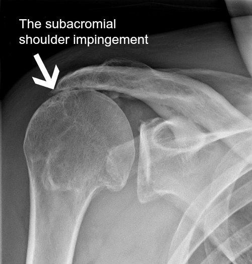
    X-ray:- On an X-ray a loss of subacromial space may be seen indicating of SAIS, but it should not be used solely for diagnosing SAIS.

    Other imagings used are US and MRI.

    Presence of a tear on MRI does not necessarily correlates with symptoms, especially in the older population (Matava, Purcell & Rudzki 2005, p. 1411).


    MANAGEMENT:-

    There is no one recipe for treating this kind of shoulder pain. Systematic Research on Exercises and manual therapy effects in Impingemnt says:-

    1. Exercises are effective in reducing pain (also stated by Senbursa, Baltaci & Atay 2007, p. 920; Trampas & Kitsios 2006, p. 125; Michener, Walsworth & Burnet 2004, p. 163)
    2. Home exercise is effective as supervised exercise.
    3. May be better effect if combined with manual therapy (also stated by DeSantis & Hasson 2006, p. 78; Trampas & Kitsios 2006, p. 127; Faber et al. 2006, p. 21; Senbursa, Baltaci & Atay 2007, p. 915; Michener, Walsworth & Burnet 2004, p. 162).
    4. Acromioplasty with postoperative exercise can improve symptoms.
    5. Functional shoulder brace may be effective in reducing pain and increase strength.
    6. Faber et al. (2006, p. 21) found that combination of exercise and manual therapy is most effective in improving function.
    7. Senbursa, Baltaci & Atay (2007, p. 915) found that improvement in function, strength and decrease in pain is significantly higher with manual therapy compare with self-training in patients with impingement syndrome. Combining manual therapy with supervised exercise may be even more beneficial (Senbursa, Baltaci & Atay 2007, p. 920).
    8. DeSantis and Hasson (2006, p. 86) found that MWM might be effective as a treatment for patients with SAIS in improving function, AROM and decreasing pain.
    9. The findings challenge the belief that posture and its concomitant muscle imbalance comprise an etiologic factor in SAIS’ (Lewis, Green & Wright 2005, p. 385).
    10. Kibler (2006, p. 42) states that there is clear link between impingement and scapular dyskinesis and thus must be addressed.

    1. Lewis, Wright and Green (2005, p. 72) found that correcting posture (scapula + thorax) with taping, increase Flexion and Abduction ROM and delayed the point at which pain was first experienced, but didn’t change pain intensity. The purpose of the tape was to extend the thoracic spine, and retract, depress and posteriorly tilt the scapula (Lewis, Wright & Green 2005, p. 82).
    2. Multiple SR (Green, Buchbinder & Hetrick 2003; Michener, Walsworth & Burnet 2004; Van Der Heijden 1997 cited in Kuhn 2009, p. 155) concluded that using US is of minor value in the treatment of shoulder pain; therefore using US is of no value for treating impingement syndrome.
    3. ‘There is no data for or against the use of cold or heat as a modality; thus, their use must be optional at this point’ (Kuhn 2009, p. 155).
    4. Cumpston et al. (2007, p. 1-2) in their SR concluded that there is some evidence that glyceryl trinitrate patches are more effective than placebo in the short term (first week of symptoms), and not clear on the long term effect. In addition, headaches were found to be common side effect in one of the studies. There is insufficient evidence to assess its effectiveness in treating rotator cuff disease (Cumpston et al, 2007, p. 15).
    5. Faber et al. (2006, p. 7) in their SR found conflicting evidence regarding improvement in function after steroid and analgesic injections.
    6. Regarding functional limitations a SR from 2006 reported ‘strong evidence that extracorporeal shock wave therapy (ESWT) is no more effective than placebo’ (Schmitt et al. 2001; Schmitt et al. 2002; Speed et al. 2002 cited in Faber et al. 2006, p. 21).
    7. Bal et al. (2009, p. 31) showed that there is no distinct advantage of using low level laser therapy (LLLT) in combination with 12 weeks home exercise program compare to exercise alone, in relation to pain and function among people with SAIS.
    8. Faber et al. (2006, p.21) in their SR found that laser is not superior to placebo in regard to functional limitations.
    9. CT evaluated the effectiveness of PEMF in the acute phase of SAIS, found that there isn’t additional benefit to PEMF in that stage (Aktas, Akgun & Cakmak 2007, p. 1234).
    10. They also found that patients that treated by both nerve block and standard rehab ( ice, pendulum, electrical stimulation, accessory joint play, strengthening, stretches etc) reported significantly less pain during physiotherapy sessions and had better final results compare with standard therapy alone.

    These 20 points on various types of treatments preferred for SA impingement or RC impingement are based on systematic research studies and are usually followed at our Physiocure once the patient is examined and diagnosed correctly.

    CONCLUSSION:-

    Rotator cuff disorders and subacromial impingement are well connected and can predispose to one another. Many theories have been suggested to try and explain the pathoaetiology of these disorders, but it is still a mystery to some extent. To date there is still a debate whether the cause is more due to extrinsic or intrinsic mechanisms, but it seems that the intrinsic theory is gaining momentum in recent years.

    Regarding management, there are many tools that can be used; none of them is a “gold standard” treatment and most of them are lacking clear clinical benefit. However, it seems that combination of treatments including some “fancy” manual therapy techniques can contribute to cope with the challenge of treating this pathology.

    Finally, if conservative approach has failed, surgical treatment can be considered.

    REFERENCES:-

    1. Aktas, I, Akgun, K & Cakmak, B 2007, ‘Therapeutic effect of pulsed electromagnetic field in conservative treatment of subacromial impingement syndrome’, Clinical Rheumatology, 26, pp. 1234-39.
    2. Bal, A, Eksioglu, E, Gurcay, E, Gulec, B, Karaahmet, O & Cakci, A 2009, ‘Low-level laser therapy in subacromial impingement syndrome’, Photomedicine and Laser Surgery, 27, no. 1, pp. 31-6.
    3. Buchbinder R, Green S, Youd JM. Corticosteroid injections for shoulder pain. Cochrane Database of Systematic Reviews 2003, Issue 1. Art. No.: CD004016. DOI: 10.1002/14651858.CD004016.
    4. Cleland, J 2007, Orthopeadic Clinical Examination: an evidence based approach for physical therapists, Saunders, Philadelphia, USA.
    5. Codsi, MJ 2007, ‘The painful shoulder: When to inject and when to refer’, Cleveland Clinic Journal of Medicine, 74, no. 7, pp. 473-88.
    6. Coghlan JA, Buchbinder R, Green S, Johnston RV, Bell SN. Surgery for rotator cuff disease. Cochrane Database of Systematic Reviews 2008, Issue 1. Art. No.: CD005619. DOI: 10.1002/14651858.CD005619.pub2.
    7. DeSantis, L & Hasson, SM 2006, ‘Use of Mobilization with Movement in the Treatment of a patient with Subacromial Impingement: A Case Report’, The Journal of Manual & Manipulative Therapy, vol. 14, no. 2, pp. 77-87
    8. Dorrestijn, O, Stevens, M, Winters, JC, van der Meer, K & Diercks, RL 2009, ‘Conservative or surgical treatment for subacromial impingement syndrome? A systematic review’, Journal of Shoulder and Elbow Surgery, 18, no. 4, pp. 652-60.
    9. Faber, E, Kuiper, JI, Burdorf, A, Miedema, HS & Verhaar, JAN 2006, ‘Treatment of Impingement Syndrome: A Systematic Review of the Effects on Functional Limitations and Return to Work’, Journal of Occupational Rehabilitation, 16, no. 1, pp. 7-25.
    10. Haahr, JP & Andersen, JH 2006, ‘Exercises may be as efficient as subacromial decompression in patients with subacromial stage II impingement: 4-8 years’ follow-up in a prospective, randomized study’, Scandinavian Journal of Rheumatology, 35, no. 3, pp. 224-8.
    11. Kibler, WB 2006, ‘Scapular Involvement in Impingement: Signs and Symptoms’, Instructional course lectures, 55, pp. 35-43.
    12. Lewis, JS 2009a, ‘Rotator cuff tendinopathy’, British Journal of Sports Medicine, vol. 43, pp. 236-41.
    13. Lewis, JS 2009b, ‘Rotator cuff tendinopathy/subacromial impingement syndrome: is it time for a new method of assessment?’, British Journal of Sports Medicine, vol. 43, pp. 259-64.
    14. Lewis, JS, Wright, C & Green, A 2005, ‘Subacromial Impingement Syndrome: The Effect of Changing Posture on Shoulder Range of Movement’, Journal of Orthopaedic & Sports Physical Therapy, 35, no. 2, pp. 72-87.
    15. Michener, LA, McClure, PW & Karduna, AR 2003, ‘Anatomical and biomechanical mechanisms of subacromial impingement syndrome’, Clinical Biomechanics, 18, pp. 369-79.
    16. Michener, LA, Walsworth, MK & Burnet, EN 2004, ‘Effectiveness of Rehabilitation for Patients with Subacromial Impingement syndrome: A Systematic Review’, Journal of Hand Therapy, vol. 17, no. 2, pp. 152-64.
    17. Senbursa, G, Baltaci, G & Atay, A 2007, Comparison of conservative treatment with and without manual physical therapy for patients with shoulder impingement syndrome: a prospective, randomized clinical trial’, Knee Surgery, Sports Traumatology, Arthroscopy, 15, no. 7, pp. 915-21.
    18. Trampas, A & Kitsios, A 2006, ‘Exercise and Manual Therapy for the Treatment of Impingement Syndrome of the Shoulder: A Systematic Review’, Physical Therapy Reviews, vol. 11, pp. 125-42.


  • Meniscal Injuries

    It’s high time a layman knows some facts about meniscal injuries that happen while playing various sports. one of them and the most important being u don’t need surgery to recover properly from a meniscal tear!!

    This Meniscal injury is common in sports such as football, rugby and other high-intensity contact sports but is also common, with no major trauma, as degenerative tears in older athletes or sedentary individuals. They occur either in isolation or combined with a ligament injury, for example, of the MCL or ACL Injury (all 3 together injured are known as the unholy triad)

    WHAT ARE MENISCI?

    • The menisci are cartilaginous structures that deepen their respective tibial articular
    • By increasing the concavity of the tibia, they play a role in stabilizing the knee. In addition, the menisci contribute to joint lubrication and nutrition.
    • The menisci have an important role as a buffer absorbing some of the forces placed through
      the knee joint, thus protecting the otherwise exposed articular surfaces from damage.
    • The medial meniscus is crescent shaped and attaches to the tibia in front of the ACL and in the intercondylar fossa at the back
    • The lateral meniscus is more circular and attaches to the tibia in front of the ACL and posteriorly behind the tibial eminence but anterior to the medial meniscus.
    • It also is attached to many other ligaments which connect the tibia and the femur within the knee joint.
    • The front portion of the menisci (known as the anterior horn) are further connected with each other via a ligament and are more stable while,
    • The back portion of both the menisci (known as the posterior horn) is connected to different structures.
    • The back portion of the medial menisci is the most susceptible to injury.
    • Thus, it is important to preserve as much of the menisci as possible after injury

    MECHANISM OF INJURY:-

    For Acute injuries:- usually startes with uncontrolled pivot in the knee or during cutting activity in any game. initailly the knee swells up and there will be pain in the innner portion of the knee. if the tear is bad then the knee might get locked up during the episode, rendering the athelete unable to play.

    TYPE OF MENISCAL TEARS:-

    • Meniscal tears can be classified as acute or degenerative.
    • Acute tears are from excessive force applied to a normal knee and meniscus.
    • This is different from a degenerative tear, which results from repetitive normal forces acting upon a worn down meniscus.
    • Tears can also be described based on pattern and location.
    • These tear patterns include vertical longitudinal, oblique, transverse (radial), horizontal, meniscal root, bucket-handle, and complex.
    • Tears can located in the avascular or vascular zone (e., white, red-white, red-red), which influences healing potential either spontaneously or after surgical repair.


    RISK FACTORS FOR MENISCAL INJURY:-


    A systematic research study showed strong evidence that age (older than 60 years), gender (male), work-related kneeling and squatting, and climbing stairs (greater than 30 flights) were risk factors for degenerative meniscal tears. They also found strong evidence that playing soccer and playing rugby were strong risk factors for acute meniscal tears. Waiting longer than 12 months between the anterior cruciate ligament injury and reconstructive surgery was a strong risk factor for a medial meniscal tear but not for a lateral meniscal tear.

    PHYSICAL EXAMINATION:-

    Usually acute knee injuries show the signs and symptoms below, however they may be symptomless in case of degenerative tears

    • Effusion,
    • Joint line tenderness,
    • Pain with squatting, and
    • A positive McMurray test.
    • Locked knee, which cannot reach full extension, may be caused by a “bucket handle” meniscal tear that has displaced centrally into the joint, blocking a full range of motion.

    INVESTIGATIONS REQUIRED:-

    Usually an X-ray and MRI are suggested. Acute isolated meniscal injuries rarely produce changes
    on plain radiograph, whereas chronic meniscal pathology may produce the Fairbank’s
    changes previously discussed. MRI imaging is useful in cases of questionable meniscal
    damage

    MANAGEMENT OF MENISCAL INJURIES:-

    An athlete with effusion and suspected meniscus injuries should be seen by an on field health care provider who usually follows the Acute injury management protocol & is further responsible for directing the athlete to an orthopaedic or a sports physio . A regular individual walking into clinics or hospitals with knee pain history indicating of degenerative meniscal tear is usually send for rehab with a physio first. At Physiocure we practice evidence based therapy. Below are lists of factor which help health care providers decide whether to send the patient for surgery or rehab.


    Factors that may indicate that conservative treatment is likely to be successful


    Factors that may indicate that surgery will be
    required
     
    ·     Symptoms develop over 24–48 hours after injury

    ·     Injury minimal or no recall of specific injury

    ·     Able to weight-bear

    ·     Minimal swelling

    ·     Full range of movement with pain only at end of range of motion

    ·     Pain on McMurray’s test only in inner range of flexion

    ·     Previous history of rapid recovery from similar injury

    ·     Early degenerative changes on plain radiographs


    ·       Severe twisting injury, athlete is unable to continue playing

    ·       Locked knee or severely restricted range of motion

    ·       Positive McMurray’s test (palpable clunk)

    ·       Pain on McMurray’s test with minimal knee flexion

    ·       Presence of associated ACL tear

    ·       Little improvement of clinical features after 3 weeks of conservative treatment


    WHAT DOES RESEARCH SAY ABOUT SELECTING TREATMENT OPTIONS?


    • Treatment options for meniscal tears fall into three broad categories; non-operative, meniscectomy or meniscal repair.
    • Selecting the most appropriate treatment for a given patient involves both patient factors (g., age, co-morbidities and compliance) and tear characteristics (e.g., location of tear/age/reducibility of tear).
    • There is evidence suggesting that degenerative tears in older patients without mechanical symptoms can be effectively treated non-operatively with a structured physical therapy programme as a first line.
    • Even if these patients later require meniscectomy they will still achieve similar functional outcomes than if they had initially been treated surgically.
    • Partial meniscectomy is suitable for symptomatic tears not amenable to repair, and can still preserve meniscal function especially when the peripheral meniscal rim is intact.
    • Meniscal repair shows 80% success at 2 years and is more suitable in younger patients with reducible tears that are peripheral (g., nearer the capsular attachment) and horizontal or longitudinal in nature.
    • However, careful patient selection and repair technique is required with good compliance to post- operative rehabilitation.(6)


    WHEN SHOULD YOU START REHABILITATION?

    Rehabilitation should commence prior to surgery. In this period it is important to:
    • reduce pain and swelling with the use of electrotherapeutic modalities and gentle range
    of motion exercises
    • maintain strength of the quadriceps, hamstrings, and hip abductor and extensor muscles
    • protect against further damage to the joint (patient may use crutches if necessary)
    • explain the surgical procedure and the postoperative rehabilitation program to the patient

    Rehabilitation principles after arthroscopic partial menisectomy are:
    • to control pain and swelling
    • to regain pain-free active range of motion
    • graduated weight-bearing
    • progressive strengthening within the available range of motion
    • progressive balance, proprioceptive, and coordination exercises
    • return to functional activities

    WHAT DOES RESEARCH SAY ABOUT REHABILITATION?

    There was wide variation in rehabilitation protocols among clinical studies. Biomechanical evidence from small cadaveric studies suggests that higher degrees of knee flexion and weight-bearing may be safe following meniscal repair and may not compromise the repair. An accelerated protocol with immediate weight-bearing at tolerance and early motion to non-weight-bearing with immobilising up to 6 weeks postoperatively is reported. Accelerated rehabilitation protocols are not associated with higher failure rates following meniscal repair.

    There are wide opinion differences on whether to start the rehabilitation and weight bearing early or late amongst the orthopaedics. Well the above systematic research study says that an accelerated rehab protocol for meniscal injuries that is improving ROM and weight bearing early is acceptable and that it is not associated with failure rates. Hence the athletes can be put through an accelerated rehab program based on their findings based on the above evidence

    Must try this exercise if u have a meniscal injury. click on this sentence!!!

    PROGNOSIS POST-SURGERY FOR MENISCAL INJURIES:-

    Surgery (partial excision, timing or meniscus suture) will allow a return to professional sports in two weeks to several months. The knee will be more vulnerable to stress at the site of meniscus excision and in the long term, 10 to 20 years, the risk of developing local osteoarthritis is significant compared to a non-injured knee


    CONTACT US FOR MENISCAL REHAB ON 9821066050 OR EMAIL US AT BOOK AN APPOINTMENT

    REFERENCES:-

    1. Clinical Sports Medicine (3rd edition- by Peter Brukner and Karim Khan with collegues
    2. The sports injuries handbook, diagnosis and management by Christofer Rolf
    3. Sports Injuires: Mechanisms, Prevention, Treatment (2nd edition) by Freddie H. Fu (Editor), David A., M.D. Stone (Editor)
    4. Risk factors for meniscal tears: a systematic review including meta-analysis.