Blog

  • Patellar Tendinopathy

    Patellar tendinopathy is a common painful, overuse disorder, often presenting with knee pain in the front portion below the knee cap area.Also commonly known as Runner’s knee or Jumper’s knee. A thorough history and physical examination are necessary, but imaging can also aid in the diagnosis. The majority of cases resolve with physiotherapy. Once patients pass the initial inflammatory phase and remain symptomatic, treatment becomes more difficult.

    Let us first try to understand where the patellar tendon is.

    On the front of the thigh a group of muscles called quadriceps are placed. These muscles form a tendon just above the knee cap (patella) which passes on and around the patella and extends to end at the shin bone (tibia). The portion below the patella is called the patella tendon

    What is the function of this tendon?

    • It connects the quadriceps muscle to the shin bone. Or it connects the thigh to the shin over the knee joint.
    • Because of its structure it can transmit the force generated by this muscle as well as absorb ground reaction forces without minimal energy loss.
    • It is flexible in nature.
    • It provides proprioception- this means it provides constant signalling of the knee position to the brain and helps the brain respond appropriately.


    What is the tendon made of?

    • It’s made of collagen which can bear the load put on the tendon & elastin component which provides some flexibility to the tendon.
    • This combination is arranged in several layers which then forms a guy rope kind of structure made of thin fibril like ropes assembled together.
    • This guy rope is further covered by thin sheet of connective tissue called paratenon which provides nutrition to the tendon.

    the more chronic or older your tendon problem will be the deeper it will reach to the tendon and may take a longer time to get the structural integrity back. but the best part is it is possible to restore the structural integrity with just physio


    How does the tendinopathy develop?

    • Tendinopathy means some pathology in the tendon.
    • When the tendon is suddenly loaded beyond its capacity to handle, there is some damage to the minute structure within the tendon. If these structures are not able to recover appropriately, overall the tendon is not fit to even take the normal load leading to tendinopathy. This would be the sudden overload theory.
    • Secondly if the tendon is repetitively loaded without sufficient time for recovery, slowly over a period of time the minute structures within the tendon are not able to take the load leading to the tendinopathy.
    • Thirdly the quadriceps muscle force generation has an effect on the tendon. Is it is tight the force generated cannot be dissipated properly or if there is continuous eccentric load which the muscle can’t handle, then the tendon takes the beating.


    The location where the tendon tends to develop a pathology?

    Usually the tendon injury in detail is identified by its location that is, whether tendon is injured at its attachment on the knee cap, in the middle part of the tendon or at the lower portion attached on the shin bone is injured. The evidence based on a research below states the following

    “This study confirms that patellar tendinopathy is not restricted to the proximal pole. Although the proximal pole was the most commonly involved location, distal pole involvement was common occurring in 38% of scans. The vast majority of previous studies have concentrated on proximal disease to the neglect of distal disease. Involvement of the mid patellar tendon was less common and when it did occur it almost always occurred in conjunction with pathology of either the proximal and/or distal poles. Isolated involvement of the middle portion of the patellar tendon was very rare (occurring in only one scan). As a result of this study the following recommendations are made:

    This means that the tendon can get affected at the bone attachment near the patella or the shin bone, however getting injured in the middle part of the tendon is rare. Also the rehab program is different depending on the location of the injured tendon.

    How does the tendon respond to loading?

    When we talk about tendon loading, what we are discussing is repetitive jumping activities or exercises where the quadriceps muscle needs to be stretched and handle the body weight at the same time which directly loads the tendon. To understand how the pathology develops it is necessary what factors affect the loading of the tendon.

    • If the tendon is thicker or longer it can with stand more load
    • With age the load bearing capacity reduces and it takes longer time to recover.
    • The tendon cannot withstand a situation where the tendon is getting compressed and loaded.
    • It cannot withstanding loading continuously especially when it is fatigued
    • If the leg has been immobilized for a long time, the tendons load bearing capacity reduces.
    • The tendons microcirculation if impaired, it will affect the nutrition to the tendon and indirectly delay the healing, in that case again the tendon may not be able respond normally to normal loads.
    • Local injection of corticosteroid into the injured tendon cause further thinning of the tendon and decreased strength, affecting the ability to bear load.
    • Diabetes affects the healing of the tendon and reduces the ability to take load.
    • Reduced oestrogen hormone can cause loading issues and lead to tendinopathy.

    Having said that , the rehab for any tendinopathy is based on loading exercises which are used in a biomechanically correct position to help reduce pain and build the tendon strength back. so don’t get confused. remember injury is caused due to faulty loading and overuse and injury will be resolved with correct loading and appropriate rest.


    What are the Risk factors for developing patellar tendinopathy?

    For nine risk factors there was some evidence:

    1. weight,
    2. body mass index,
    3. waist-to-hip ratio,
    4. leg-length difference,
    5. arch height of the foot,
    6. quadriceps flexibility,
    7. hamstring flexibility,
    8. quadriceps strength and
    9. vertical jump performance.

    Based on the present evidence, reducing body weight, increasing upper-leg flexibility and quadriceps strength and the use of orthotics may be beneficial treatment options. However, it should be stressed that the evidence for the nine identified risk factors was only limited.

    (more…)

  • Patellofemoral Pain Syndrome

    Pain in front portion of the knee, in and around the knee cap (Patella) is known as Anterior Knee Pain. This is a broad term which includes number of diagnosis like Patello Femoral Pain Syndrome (PFPS) and patellar tendinopathy. As per research around 20-40% knee injuires are reported in this area while playing any kind of sports.

    Distinguishing between PFPS and Patellar Tendinopathy as a cause of front knee pain
    can be tricky as their signs and symptoms are almost similar and required clinical experience

    ANATOMY OF THE KNEE CAP:-

    The bones of the knee and the leg include the femur, which is the large thigh bone; the tibia and fibula, which are the leg bones between the knee and ankle; and the patella, which is known as the kneecap.

    The patella is lined with cartilage which is actually the thickest cartilage in the whole body due to the massive forces that go through it e.g. the force going through the kneecap when coming downstairs is 3.5x bodyweight.

    The quadriceps muscles are placed in front of the thigh which controls the patella movement. They insert on the shin bone as patella tendon which acts as a pulley in directing the direction of forces on the patella.

    The most important function of the patella is its role in facilitating the extension of the knee by increasing the efficacy of the quadriceps muscle.

    how does it move on the knee?

    The knee cap , when your leg is straight sits on the outer part of the knee. When the knee is extended, the tightened quadriceps pulls the patella upwards until the upper border reaches beyond the femoral trochlea groove As the knee starts bending , the knee cap moves horizontally inwards.


    Because its shape is triangular on the inside where the cartilage is and the space wher it sits is also a triangular sort of cave, it can move from out to in every time you bend you knee.

    The Quadriceps mainly control this side to side & up and down movement of the patella. If we try to go in details quadriceps is a set of 4 muscles, of the these there is Vastus medialis muscle (VM) on the innerside fo the thigh and, Vastus lateralis muscle (VL) on the outer side of the thigh.

    For the knee cap to smoothly move from side to side, both these muscles need to have a balance of strength and flexibility which could be another reason for your PFPS. Another fact is the muscle on the outer side that is vastus lateralis is anatomically stronger than the inner one and hence the chances of imbalance are frequent.

    How much weight does it take?

    When you are walking it take around 0.5kgs of weight, while jogging the weight is around 7-8 times your body weight. How does it affect your knee pain is the question.

    If you look at the picture showing arrows where the force is directed on the knee cap when moving it shows that the knee cap is under maximal pressure when you knee is bent up till 90 degrees. Hence it is common to experience pain while doing activities where knee is bent when you have PFPS.

    Gender differences

    Women have smaller knees and hence shorter patellar tendon moment arms than men. Subsequently the PRF increases by up to 20% for the same knee extending moment which would explain the somewhat higher frequency of patello-femoral disorders in females.

    SIGNS AND SYMPTOMS:-

    • Although it may be difficult for the patient with anterior knee pain to be specific, the area of pain often gives an important clue as
      fat pad impingement

      to which structure is contributing to the pain. For example, pain at the back of patella (knee cap) suggests PFPS, while pain at the lower tip of the knee cap suggests pinching of the fat pad present in that area.

    • The type of activity could also hint onto the diagnosis like for example activities a sports person complaining of pain at the knee cap who is involved in repetitive jumping like basketball, volleyball usually load the patella tendon, so it could be more of a tendinopathy while the same person who has been running could be more of a PFPS. Hence it is important for the patient to describe his activities or sport in detail.
    • How the pain started is also important. The pain for PFPS usually has a unnoticed episode of knee pain earlier like falling on the knees, previous knee ligament injury or post surgery, recurrent ankle sprains etc
    • The pain usually get worst on loading the knee like climbing, walking, running or for regular people it could also get worst with sitting for a long time.

    • Another differentiating feature is usually the pain reduces after warm up and returns after the body cools down in athletes then it’s the tendon which troubled and not the knee cap
    • Recurrent crepitus is suggestive PFPS but the knee cap moving too much out of position could be more of an instability issue
    • Another common symptom is giving way of the knee, but that could be due to PFPS or acl injury, meniscal injury, patella dislocation. So its an important sign but needs further understanding clinically.
    • Last but not the least, PFPS usually the swelling is more on one corner of front of the knee, but a fully swollen knee could suggest more of a knee joint problem. However both could co-exist together.

    THE KINEMATIC CHAIN AND THE RELATION BETWEEN CORE/HIP/KNEE STABILITY:-

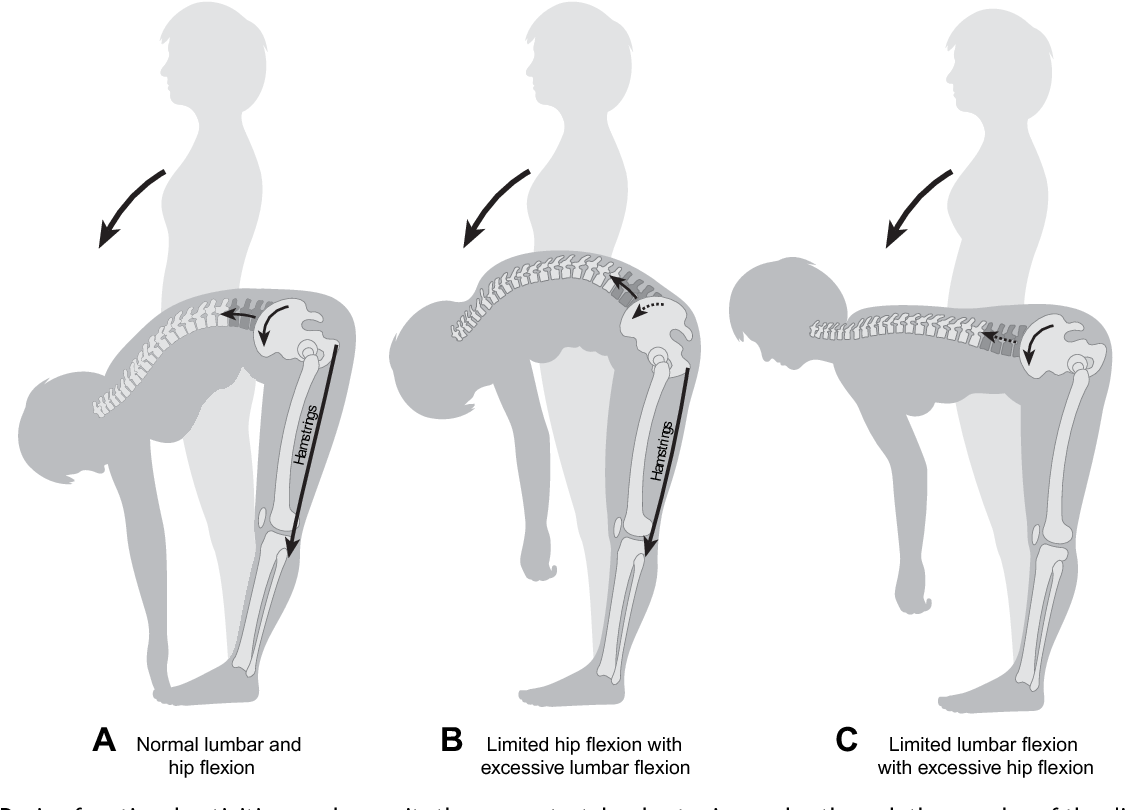
    From a sports physio point of view, we look at the biomechanical causes of your knee pain. Most of the research has indicated that stability at the hip and knee are inter-related and that a weakness/tightness in hip or back muscles could indirectly influence you knee pain. Hence most of the rehab protocols focus on improving the hip and back/core strength along with knee.

    The other reason to do this is that our body musculoskeletal structure actually functions in kinematic chains which helps us perform these complex movements. So it would be logically sound to work on the chain than just the area of the pain.

    For example:- have you heard about this structure called IT band. Its pretty famous with the fitness and sports guys with information. Now most of the times if you have been diagnosed PFPS your health care provider may work on the IT band along with the knee, but if you approach this problem from the kinematic chain theory it could reveal a weakness/tightness anywhere in the chain. It may be your foot or hip/ spine or the IT band. Hence evaluating this pain functionally leads to solution of the problem to the core and not just on the top.

    MANAGEMENT OF PFPS:-

    Usually the treatment is based on following principals:-

    • Reduce pain
    • Restore mobility and stability
    • Correct biomechanics

    References

    1. Anterior knee pain: clinical Sports medicine- peter Brukner and karim khan (3rd edition)
    2. Basic kinematics and biomechanics of the patello-femoral joint. Part 1: The native patella.
      1. Oliver S. Schindler, William N Scott
      2. Published 2011 in Acta orthopaedica Belgica
      3. https://www.semanticscholar.org/paper/Basic-kinematics-and-biomechanics-of-the-joint.-1%3A-Schindler-Scott/477b76e5860204ec747c5ea87c34e9e6633fa793


  • Running

    Running is one of the most popular sports in the world. This not only covers marathons and track and field running – running is also part of the training in virtually all other sports, including soccer, rugby, handball and triathlon. Modified levels and intensities of running can also be used as alternative training for most athletes with certain types of injuries

    Running is a very efficient method for maintaining or improving overall fitness and increasing the structural strength of the leg muscles. Running can be over a distance, in a variety of intervals or as a varied running-jogging-walking program. The intensity and the distance must be proportionate to the runner’s ability and objectives.

    Running on a hard surface

    However,before running on a hard surface, such as roads or astro turf, the runner must be able to withstand the repetitive impact of the ground on to the legs, as each stride will create an impact force of five to ten times body weight for a fraction of a second. Since each stride stresses the same structures, their tensile strength and endurance lie between positive training effects and injury.

    Running on a hard surface mainly loads the lower limbs, where 90 per cent of running injuries are found. The most common error an inexperienced runner makes is to run too fast and too long too early, so that training causes new injuries instead of promoting the healing of another.

    Prescribing hard surface running

    It is important to use common sense when prescribing hard-surface running. For an inexperienced runner the safest way to build up performance after an injury is a slow and steady running tempo,
    including a proper warm-up. The best test of improvement in running capacity is repeatedly to measure the runner’s effort and time in a simple test race.

    Running for general fitness

    For a fairly unfit but otherwise healthy person who wants to improve their general fitness and aerobic performance, running can be recommended as part of a progressive program. From a reasonable starting point, such a program would usually increase less than 15 per cent in distance and intensity per year.

    When to stop/reduce running on a hard surface!!

    • An elite marathon runner with an over-use injury may simply reduce their running time from two hours to one hour per day; not pushing over the pain threshold but gradually increasing the time day by day.
    • A 130 kg rugby player, even though extremely fit, is not a good candidate for long-distance or road running. Their knees will undoubtedly say ‘no!’ to this madness.
    • This type of exercise also cannot be recommended for obese or generally unfit recreational athletes or people with structural knee or hip problems, such as osteoarthritis.


    How do you know your running capacity is decent!!

    A reasonable running tempo that can be maintained for 30 minutes is essential for a persistent training effect. Runners should aim for a pace at which they can chat with a running mate while breathing almost normally; this is equivalent to 60 to 70 per cent of maximum aerobic capacity.

    The subjective experience of running is far more important than the heart frequency, which is not directly proportionate to the runner’s feeling. Even with the same heart frequency, for example 160 beats per minute, running can be very easy one day and very uncomfortable the next.

    Training to run efficiently

    The subjective experience of training is very important for a non-runner’s motivation. Since the surface is consistent for each stride the same structures in the lower limbs will be put under repeated stress. While this leads to a functional adaption of the strength of the structures it can also lead to
    injuries in the short term.

    The comfortable fitting shoe for running

    This also highlights the importance of proper, comfortably fitting of The Running Shoe with a cushioned sole, which can reduce the impact from touchdown in the stride, distribute the forces and provide stability to the ankle and foot.


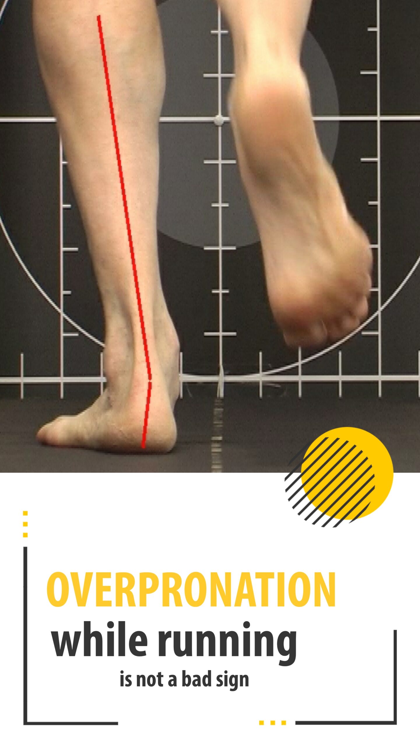
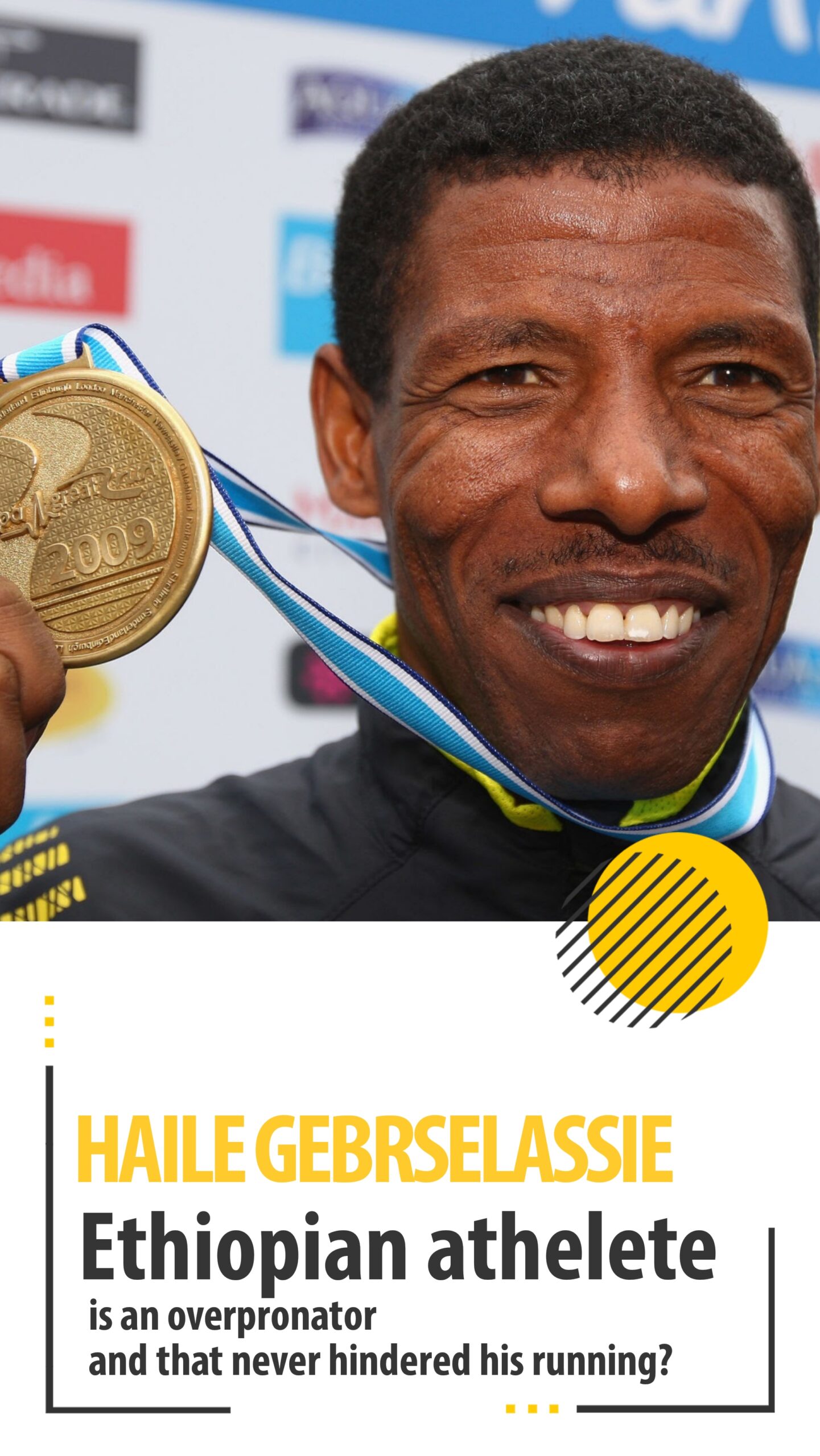
    overpronation is normalmarketing strategyEthopian athlete


    Regular runners will soon find their own style, but might have more trouble getting a consistent style of footwear. Most running injuries are caused by training errors.

    We usually recomment these ankle mobility exercises for runners training for a marathon

    Don’t get carried away!!

    The commercial running shoe market is unfortunately mainly fashion-orientated and new models are pumped out every six months. Despite improved bio mechanical knowledge, which manufacturers claim has revolutionized the market, and lighter high-quality materials and technology within the sole to compensate for different individual factors, modern running shoes do not last long. It may also be questioned whether they have reduced the incidence of injuries.

    Soft Surface Running

    Running on soft surfaces maintains or increases fitness and tensile strength of the lower limbs and with less eccentric impact compared to running on hard surfaces. A well-balanced running programme over beachess, in parklands or in forests can be recommended as a primary alternative
    training for most runners and other athletes with over-use injuries, even those of the lower limbs.

    People with mild or moderate knee or hip osteo-arthritis, who struggle to run 500 m on the road, may be able to jog a 5 km orienteering course without adverse effects.

    people with knee or hip arthritis must do these stretches before going for the run

    Advantages of soft surface running

    • A varied and soft running surface creates a lower impact on the musculo-skeletal system, due to the longer time for shock absorption from each stride and the wider distribution of forces over the kinetic chain.
    • On the other hand, the runner is forced to work harder, from a muscular point of view; they need to lift the knees higher when running uphill or over obstacles on the ground like vegetation.
    • This consumes more energy and, all in all, uses more muscle groups than running on hard surfaces.
    • Running in forests consumes up to twice as much energy per kilometer as road running.
    • Running on soft surfaces is less demanding for the lower limbs but, due to the increased energy demand, puts more stress on the cardiovascular and respiratory system and so is good for weight reduction and general fitness.

    Risks of soft surface running!!

    • There are risks in the prescription of this type of training. Over uneven terrain, the risk of ankle sprains and falls increases.
    • Even though temporarily running on soft surfaces can be recommended for a marathon runner with over-use injuries of the lower limbs, they must be aware of the increased risks.
    • Proprioceptive ankle training and core stability training must accompany alterations in running type.


    Who should avoid running on soft surfaces

    Older athletes, or fragile patients with osteoporosis or disabling injuries, may jog or walk on softer surfaces instead of running

    Last but not the least if you are into running you must follow these three stretches everyday to prevent yourself from injuries.

  • The Running Shoe ?

    As more and more people resort to running for health and fitness, so have the number of  Running injuries increased. A visit to Physiocure we can help you educate with the foot types, shoe designs and a simple examination can reveal which foot type you run with and is your current running shoe good enough.

    When we happened to interact with our professional runners on how they choose their shoes, we were not surprised at all. Most consumers are nowadays visiting the sports shoe showrooms and are made to walk on the treadmill, a foot scanning is done and then the shoe is prescribed at least in metro cities in India, for smaller cities it’s more of what feels comfortable and what fits my budget.

    A lot of people pick up running shoes based on

    • available discounts,
    • latest styles or
    • they are consulted by so called “storeroom shoe prescription experts” into buying the motion controlled or pronation control type footwear.

    this is common and is not always bad, but really it is a matter of concern. after 12 years of practice, I believe, a low arch, motion-controlled shoe, a high arch are secondary factors that contribute to running injuries, and its just a “fear-based marketing approach” to sell new, highly-priced running shoes

    overpronation is normalmarketing strategyEthopian athlete


    Before you go for the new shoe selection trip to these stores again, ask yourself these questions:

    • Would you take the advice of a person trained by a marketing expert into explaining the foot scanning in these stores or your physio/health care provider?
    • Did you check your foot type from a podiatrist/physio/health care provider or the sales guy at these stores?
    • Has choosing shoes on based of feel, prevented your number of sports injuries.
    • Is you running pattern in sync with the motion control shoes you’ve been offered.
    • Is the sales guy really a shoe prescription expert?
    • Was there something wrong with my last shoe or my running style or fitness?


    What does research say??

    Where can we find the truth, as physiotherapists? Peer-reviewed research is a fantastic source of information and can guide us to the truth when it comes to what Running shoe is best for our patients.

    It was once believed that those with low arches should wear “motion-control” shoes which are designed to be more rigid. Those with a more neutral or “normal” arch should be placed in “stability” shoes, and those with a high arch should be placed in more cushioned, “neutral” shoes.

    Despite common “knowledge” in the running community, selecting a shoe based on arch-height does not lead to reduced injury (1).

    Another misnomer commonly used for shoe prescription indicates that excessive pronation of the foot and ankle leads to injury, so shoes should be fitted to reduce pronation.

    Current research is inconsistent and conflicting but has not shown a strong correlation between “excessive” pronation and injury (2, 3).

    Pronation or foot posture can not confidently be used as an indicator for shoe prescription, barring further research.

    What about “minimalist” shoes?

    Once again, not enough research has been completed to confidently say that wearing minimalist shoes leads to reduced injuries. Minimalist shoes are designed to mimic running barefoot, resulting in a foot-strike closer to forefoot (4). This “strike-pattern” is linked to reduced forces placed through the shin bone ( a.k.a thetibia) and the knee.


    Also, running barefoot actually has been shown to reduce ground-reaction forces through the leg, when compared to running with neutral shoes (5).

    As discussed, much of the “common knowledge” about Running shoe selection is either false or has little evidence to support it. So, what is the best way to recommend shoes?

    (more…)

  • Training For The Mumbai Marathon….Try Deep Water Running

    With Mumbai marathon scheduled in January of each year, all those who’ve been taking up training for professionals for the marathon, here’s a different way of treating if you are injured or recovering if you think the training has gone too intense.

    Individuals who perform land based training exercises such as running or jogging, may be required to discontinue that activity if an injury occurs which may require discontinuing running for 4- 6 weeks. Runners fear such a break may lead to a decrease in fitness or an increase in body weight, and few are willing to endure long periods of inactivity. A 4-6 week period of inactivity will lead to a 14- 16% reduction in lung capacity over the 4- 6 week period. Thus, an aquatic based therapy for “active rest” from an injury. The goal is to maintain cardiovascular endurance, mobility strength and flexibility while resting the injury.

    Deep water running (DWR) consist of simulated running in the deep end of the pool aided by a floatation device (vest or belt) that maintains the head above water. The athlete may be held in one location by a tether cord, essentially running in place or may actually run through the water the length or width of the pool.

    • During DWR, the body is tilted slightly forward approximately 5 degrees past vertical, with spine in neutral position, the bend should occur at hips, not the waist.
    • The head is held comfortably out of the water, facing forward; avoid neck extension.
    • The arm action occurs primarily at shoulder and with hands relaxed but slightly closed.
    • No contact is made with the bottom of the pool, thus eliminating impact.
    • This form of running in the water closely follows the pattern used on land. However , the center of gravity on land is at hips. In water, the center of buoyancy is at lungs. To get used to this change, the athlete must retrain the body to use abdominal muscles to maintain the correct vertical posture.

    Advantages

    • Land based runners who water train maximize spped gains and these gains can transfer to land performance.
    • Water’s buoyancy eliminates the effect of gravity supporting 90% of body’s weight thereby reducing the impact and creates greater flexibility.
    • Using variable depths is very useful when recovering from an injury or after a hard training session or to partially unload the body.

    Rehabilitation protocol for an athlete with with a leg injury

    Week 1 – correct form of running used in DWR FOR 20-40 at a steady pace for 3-4 times a week. Also, exercises related to the injury

    Week 2-3 – start with cadence interval training

    Week 4 – add resistive equipments as tolerated (gloves, delta bells, aqua runners). 2 times a week interval training and 2 times a week easy running (30-45 min) with resistive equipment.

    Week 5-6 – athlete is trained in the water specifically to their sport

    For example-marathoners

    • 1 dy/wk: long run for 1-2 hrs ( depending on fitness level and timing of training)
    • 1 day/wk: interval training
    • 1 day/wk: strength run; a steady run of 20-40 min
    • 2 days/wk: easy running in the water; 30-60 min

    Use the easy days between the hard training session. It gives the body a chance to recover. The hard easy system of training works the best.

    Alternatively with regular training protocols for a particular type of marathon (half, full) which may be strenuous, deep water running can be substituted for a twice a week session.

  • Bad Posture Vs Good Posture

    Good posture is a good habit that contributes to the wellbeing of the individual. The structure and function of the body provide the potential for attaining and maintaining good posture.
    Conversely, Bad posture is a bad habit and, unfortunately, is all too common . Postural faults have their origin in the misuse of the capacities provided by the body, not in the structure and function of the normal body.(1)

    If faulty posture were merely an aesthetic problem, the concerns about it might be limited to those regarding appearance. However, postural faults that persist can give rise to discomfort, pain, or disability . The range of effects, from discomfort to incapacitating disability, is often related to the severity and persistence of the faults.(1)

    The high incidence of postural faults in adults is related to this tendency toward a highly specialized or repetitive pattern of activity. Correction of the existing conditions depends on understanding the underlying influences and implementing a program of positive and preventive educational measures. Both require an understanding of the mechanics of the body and its response to the stresses and strains imposed on it.(1)

    What does research say about this???

    1. Ten trials with 2745 participants were included in this review.The present review found low quality evidence that those who received workplace interventions did not get more pain relief than those who received no interventions.(4)
    2. There is evidence which is limited in quality to indicate that ergonomic workplace interventions can improve gross sitting posture.(5)

    However research stated that workplace interventions for correcting faulty postures have been ineffective and have very low quality or limited evidence that having a ergonomically perfect chair will solve the problem.

    A problem arises, however, because skilled performance in a variety of sport, dance, and acrobatic activities requires excessive flexibility and muscle length. Although “the more, the better” may apply to improving the skill of performance, it may adversely affect the well-being of the performer. Hence managing postural faults for athletes, dancers and active individuals in a different ball game.


    The following definition of posture was included in a report by the Posture Committee of the American Academy of Orthopedic Surgeons (6). It is so well stated that it bears repeating.(1)

    “Posture is usually defined as the relative arrangement of the parts of the body. Good posture is that state of muscular and skeletal balance which protects die supporting structures of the body against injury or progressive deformity, irrespective of the attitude (erect, lying, squatting, or stooping) in which these structures are working or resting. Under such conditions the muscles will function most efficiently and the optimum positions are afforded for the thoracic and abdominal organs. Poor posture is a faulty relationship of the various parts of the body which produces increased strain on the supporting structures and in which there is less efficient balance of the body over
    its base of support.

    FAULTY POSTURE:-

    If incorrect postures become a habit at an early age, individuals maintaining those postures may adapt and consider them comfortable, and this can cause strain on the spine, pelvis, muscles, tendons, joints, bones, and discs, which can lead to fatigue and deformation.(2)

    Thus, incorrect habits, such as excessive use of computers, use of desks and chairs without proper height, lack of health care education, lack of exercise, carrying heavy school bags, and inappropriate postures when studying or watching television, affect the shape of muscles, deform the skeleton, and cause abnormal development, which prohibit the maintenance of correct posture(1)

    When discussing pain in relation to postural faults, questions are often asked about why many cases of faulty posture exist without symptoms of pain, and why seemingly mild postural defects give rise to symptoms of mechanical and muscular strain.

    The answer to both depends on the constancy of the fault. An interesting concept which has not been mentioned most texts is the difference between a faulty posture and postural stresses  has been discussed in an interesting microblog.

    What are postural stresses?

     “Postural stress” is a challenge to your posture that is imposed on you, as opposed to something you’re doing to yourself out of laziness. Postural stress is easier to define, maybe easier to causally link to pain, and certainly often much easier to solve than poor posture. They are also the reason for most of the musculoskeletal problems arising in the name of faulty posture.(2)

    Usually with a postural stress the muscles give way because they are tired and irritated of working in that awkward position as compared to faulty posture where muscles are not used at all and eventually they tend to switch off.

    For example prolonged sitting caused your butt muscles to stop working efficiently but prolonged sitting without back support causes your neck muscles to be tired and irritated leading to poker chin posture which eventually causes neck pain.

    Postural stress and faulty posture may have many common developmental factors but it is obvious that postural stresses are situaltional where the person may not have a choice and adopting faulty posture may be out of just laziness and a foolish choice.

    For example:-

    1. Trying to sleep where it is impossible to do so, like in a aeroplane or a car seat is a postural stress whereas sleeping with 2-3 pillows slouching on the bed is a adopted foolish faulty posture
    2. Another example would be a nurse, who has to constantly bend from the back to lift patients/ perform her duties and be on toes for major part of the duty hours. This makes them prevalent from back pain and heel pain out of postural stresses. But the same nurse who chooses to wear heels everyday at work is adopting a faulty foot posture which may lead to her back pain and heel pain.
    3. Similarly an office employee who travels with the laptop for marketing may not find an ergonomically suited desk and works in the available position placing the laptop on the lap, or a table of the available height. This postural stress might contribute to neck pain or mid back pain.

    POSTURE ANALYSIS AT PHYSIOCURE:-

    For posture examination, first a basic posture is examines in standing position followed by sitting and / or supine if required. For each profession further specific examination may be added.

    For example :-

    • For a lab technician the pinches and grip holding position and the lab setup is examined.
    • For a marathon runner, the running form is examined.

    Check out our basic posture analysis form.

    Basic Posture analysis form

    MANAGEMENT OF POSTURAL PROBLEMS:-

    Inherent in the concept of good body mechanics are the inseparable qualities of alignment and muscle balance. Examination and treatment procedures are directed toward restoration and preservation of good body mechanics in posture and movement.

    Therapeutic exercises to strengthen weak muscles and to stretch tight muscles are the chief means by which muscle balance is restored.

    Good body mechanics requires that range of joint motion be adequate but not excessive. Normal flexibility is an attribute; excessive flexibility is not. A basic principle regarding joint movements can be summarized as follows:

    the more flexibility, the less stability; the more stability, the less flexibility.

    here are some simple exercises to correct a faulty upperbody posture

    What does research say?

    Research studies show that steady exercise corrects posture, which improves the balance of the body, and relaxes the whole body, which relieves musculoskeletal pain. Therefore, the development and introduction of suitable exercise programs will contribute to the physical and mental health of society.(3)

    We at Physiocure do not advise you to correct your static posture but examine you thoroughly for the postural stresses as well and help improve aches and pains arising out of these postural stresses, which eventually help in improving your faulty posture.

    REFERENCES:-

    1. Muscles Testing and Function with Posture and Pain (5th Edition) Florence Peterson Kendall, Elizabeth Kendall McCreary, Patricia Gelse Provance, Mary, Mclntyre Rodgers, William Anthony Romani.
    2. Poor posture versus postural stress –  www.painscience.com/microblog.html
    3. Effect of an exercise program for posture correction on musculoskeletal pain  DeokJu Kim,1 MiLim Cho,2 YunHee Park,3 and YeongAe Yang4,
    4. Workplace interventions for workers with neck pain Aas RW, Tuntland H, Holte KA, Røe C, Lund T, Marklund S, Moller A
    5. Workplace interventions to improve sitting posture: A systematic review.Swinton PA1Cooper K2Hancock E3.Prev Med. 2017 Aug;101:204-212. doi: 10.1016/j.ypmed.2017.06.023. Epub 2017 Jun https://www.ncbi.nlm.nih.gov/pubmed/28647545
  • Headache


    Headache

    A physiotherapists approach to headache has been suppressed or ignored for long . in this article I intend to educate you how u can solve your headache or vertigo issue just with physiotherapy

    Headache is a symptom of diseases or a disorder of many origins in the body including the brain, the wiring (nerves), vessels carrying blood, eyes, ears, sinuses, nose and throat, dental and psychiatric as well. Successful management will only be achieved by a successful diagnosis.

    The reason that differentiation is often a difficult process is because the health care provider must rely on the nature and pattern of  headache and furthermore the symptoms of different causes of headache overlap or the patient may be suffering from more than one form of headache simultaneously.

    In such cases, it would be necessary to distinguish between types of headache and determine how much of a role the structures of the cervical spine have in the cause of the headache.

    There are 6 different types of headaches:-

    1. Cervicogenic Headache
    2. Tension Headache
    3. Migraine
    4. Cluster Headache
    5. Neural Headache
    6. Post traumatic Headache

    Physiotherapy can help resolve all those headaches to some extent and can provide a complete cure to cervicogenic headache.

    Symptoms:-

    What findings would you expect for cervicogenic headaches?
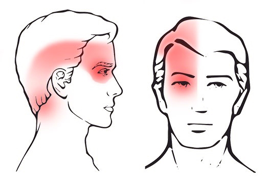
    1. Area:
    – frontal, occipital, retro-orbital, temporal
    – there is often associated neck pain (sub-occipital)
    – symptoms usually start in neck, sub-occipital or occipital
    – they are usually unilateral, may be bilateral but does not change sides during attacks. (ie. Starts on one side then spreads to the other)
    2. Quality:
    – an ache or dull boring pain is the most common description
    – can have shooting pain (deep)
    – moderate to severe level
    3. Associated Symptoms:
    – nausea, vomiting and photophobia (ipsilateral to side of pain)
    – dizziness
    – light-headedness
    – visual disturbances
    – tinnitus
    4. Frequency and duration:
    – can be episodic (few hours to few days) or chronic (semi-continuous
    or 2-3 per week
    5. Time and Mode of onset:
    – may be present upon waking and may worsen as day goes on/
    activity dependent
    6. Aggravating and relieving factors:
    – sustained neck postures or movements
    – may not know particular pattern
    -stress or tension may increase headache
    7. General Medical History:
    – no family history
    – more common from 20-50 age group
    – females > males
    8. History of onset:
    – trauma or degenerative joint disease of upper cervical joints

    Management:-

    The Physio treatment is focused on the following goals:-

    • Improve neck/ upper back mobility
    • Improve neural mobility
    • Correct muscle imbalances around cervical/shoulder/shoulder blade
    • Improve posture

    What research says about effect of physio treatments on different kind of headaches:

    1. There were no differences in headache-related and demographic characteristics between the groups at baseline. The loss to follow-up evaluation was 3.5%. At the 12-month follow-up assessment, both manipulative therapy and specific exercise had significantly reduced headache frequency and intensity, and the neck pain and effects were maintained (P < 0.05 for all). The combined therapies was not significantly superior to either therapy alone, but 10% more patients gained relief with the combination. Effect sizes were at least moderate and clinically relevant

    It means that for cervicogenic headaches the physio treatment both manipulations and /or exercises were effective . they were helpful not only for short term relief but for long term as well

    1. Migraine research review- For the prophylactic treatment of migraine headache, there is evidence from a high-quality study that spinal manipulation may be an effective treatment option with a short-term effect similar to that of a commonly used, effective drug (amitriptyline). There were fewer side effects associated with spinal manipulation.

    It means that for short term relief of migraine you can also resort to physio treaments instead of medications.

    1. For the prophylactic treatment of cervicogenic headache, there is evidence from a high-quality study that both neck exercise (low intensity endurance training) and spinal manipulation are effective in reducing headache intensity and frequency in the short and long term in comparison to no treatment. Except for reduction in headache duration, there is no advantage to combining the two therapies. From two more high-quality studies there is evidence that spinal manipulation is effective in the short term in improving pain and other secondary headache outcomes in comparison to massage or placebo spinal manipulation. Weaker evidence from a lower-quality study showed that spinal manipulation was more effective for pain reduction in the short term than spinal mobilization or no treatment.

    It means that specific and low level physio treatments can be used to prevent development of such cervicogenic headaches. The effects are much superior to just massage and/ or chiropractic treatments.

    1. There is some evidence that spinal mobilization may have better short-term reduction in pain intensity than cold packs in the treatment of post-traumatic headache.

    It means that manual therapy is more effective than just cold packs in a headache caused due to some kind of trauma.

    Exercises for cervicogenic headache

    Although most clients require hands on treatment, in the wake of pandemic we have posted few simple exercises to relieve headache on our youtube channel. try these!!

    Conclusion:-

    Headaches of cervical origin, migraines, neural type as well as post traumatic types can be benefited by Physiotherapy.

    References

    1. A Randomized Controlled Trial of Exercise and Manipulative Therapy for Cervicogenic Headache

    Jull, Gwendolen, PT, PhD,*; Trott, Patricia, PT, MSc,†; Potter, Helen, PT, MSc,‡; Zito, Guy, PT, Grad Dip Manip Ther; Niere, Ken, PT, Mph,; Shirley, Debra, PT, BSc; Emberson, Jonathan, MSc,#; Marschner, Ian, PhD,#; Richardson, Carolyn, PT, PhD*

    Spine: September 1, 2002 – Volume 27 – Issue 17 – p 1835-1843 https://journals.lww.com/spinejournal/Abstract/2002/09010/A_Randomized_Controlled_Trial_of_Exercise_and.4.aspx

    1. Non-invasive physical treatments for chronic/recurrent headache

    Gert Brønfort1 , Niels Nilsson2 , Mitchell Haas3 , Roni L Evans1 , Charles H Goldsmith4 , Willem JJ Assendelft5 , Lex M Bouter6

    http://dare.ubvu.vu.nl/bitstream/handle/1871/27456/271236.pdf?sequence=2

    1. Cervical Headache: A Review. In: Grieve: Grieve’s Modern Manual Therapy of the Vertebral Column. Jull G.A. (1994)


  • Movement Based Therapy

    movement impaitments

    What is this movement based therapy?

    Its 2020, we all are partly or completely urbanized and clearly we all need some guidance on how to move. Now thats where physio’s can play a crucial part apart from various other exercise science experts.

    Let me tell you how!!

    There are a lot of physio’s who can help solve your aches and pains by just advising the correct movements. like i said its 2020!  Have you heard of therapies to solve your problems purely based on movement corrections. we at physiocure practice few of them.

    Being into sports physio, I try to follow a mix of these various movement therapies to help all my clients with their injuries like Lowback pain, Headaches, neck pains shoulder impingements, knee pains, faulty postures etc

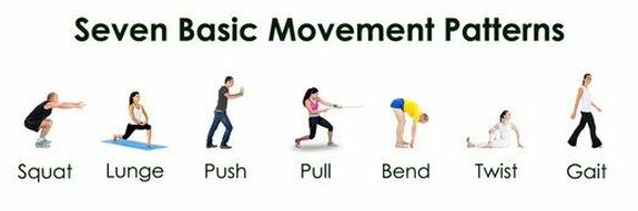
    These are based on functional movement screenings for example to check how are you moving in the 7 basic movements of human life.

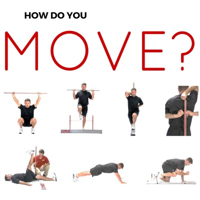
    This therapy is not just restricted to treating your aches and pains. we as physiotherapists can also guide you and teach you correct way to do gym exercises by just examining once as to How do you move?


    Its called Functional movement screening. its very effective for active people as well as athletes.

    For clients who are experiencing lower back pain or neck pain, I usually follow a therapy based on Dr Shirley sahrmanns Movement Impairments Syndromes. It has been so far very effective in treating more than 10,000 clients at the clinic for various spine problems in the past decade.

    What is Movement Impairment syndrome (MIS) ?

    In 2000, Shirley Sahrmann, a renowned physiotherapist  along with her colleagues introduced a new school of thought which uses examination of movements and identifying impairments which eventually help physiotherapists design treatment programs to treat musculoskeletal conditons like low back pain, neck pain, shoulder pain and  many more. The basic concepts of MIS are based on the following points.

    • Musculoskeletal pain syndromes are the result of cumulative micro-trauma from accumulation of tissue stress and irritation resulting from sustained alignments or repeated movements in a specific direction(s) associated with daily activities.
    •  the reason certain musculoskeletal pains develop because there has been minute trauma/stress on that tissue occurring for over years or a long time due to faulty adopted positions for your work or leisure.
    • The joint(s) that is moving too readily in a specific direction is the site of pain generation.

    As a result, the affected area will move excessively in a particular direction, for example a person who has been using a computer with a wrong ergonomic setup and adopting wrong postures for years will be able to poke his chin out much more than an individual who has just started and complain of occasional neck pain.

    • The readiness of a joint to move in a specific direction, i.e., the micro-instability, combined with relative stiffness, the neuromuscular activation pattern and motor learning contribute to development and persistence of the path of least resistance.
    •  The reason a joint can be moved excessively in one direction is the structures supporting it tend to stretch out and cannot be restored to normal length, the muscles surrounding that joint learn to function in a faulty manner and it ends up becoming the new normal position for that joint.
    • Treatment is based on correcting the impaired alignments and movements contributing to tissue irritation as well as correcting the tissue adaptations, such as relative stiffness, muscle weakness, and neuromuscular activation patterns.
    •  Once your physio is able to identify this impairment, the treatment is as simple as correcting that impairment via corrective exercises and allowing the surrounding muscles and tissues to adapt to a better normal position.
    • Training to correct impaired alignments and movements instead of training “isolated muscles” will induce appropriate neural and musculoskeletal adaptations.
    • In summary, using this concept, the faulty movement is tested and corrected than typical physiotherapy/orthopaedic treatment and indirectly /directly the ailment is resolved with appropriate and simple measures.

    The following example illustrates how correcting the impaired alignments and movements address the cause of the pain, which is not achieved by identifying the pathoanatomical source of the symptoms.

    • A patient is referred to physical therapy with the diagnosis of Supraspinatus Tendinopathy.
    • Tendinopathy is the pathoanatomic source of pain.
    • After assessing the patient’s scapular and humeral alignments and movements and associated symptom behaviour the physical therapist makes a diagnosis of insufficient scapular upward rotation with humeral anterior glide- basically the physio concludes that the shoulder blade is the reason of his pain and its inefficient work needs to be corrected.
    • The other components of the examination identify the contributing factors that include (1) relative stiffness, (2) muscle strength, and (3) neuromuscular activation patterns.
    • The idea behind the KPM is that classifying the patient according to impaired alignments and movements (i.e., Scapular Insufficient Upward Rotation, Humeral Anterior glide) is more useful to guide physical therapy treatment than identifying a pathoanatomical problem because these are the impairments to be corrected

    check out this advanced shoulder blade exercise we recommend to our clients

    TREATMENT PRINCIPALS:-

    • Treatment is based on correcting the impaired alignment and movement patterns as well as correcting the tissue adaptations associated with the impaired alignment and movement patterns.
    • Treatment includes patient education, analysis and correction of daily activities and prescription of specific exercises.12798081
    • Patient education refers to educating the patient about how the repetition of impaired movements and sustained alignments in a specific direction may be related to his musculoskeletal condition and how to correct the impairments during all of his daily activities, particularly those that cause symptoms.

    For example, patients with Scapular Depression Syndrome may be taught to keep their scapula elevated by supporting their arms while working at a computer. The goal of the support is to reduce the sustained load on the cervical spine and the muscles that elevate the scapulae

    • The specific exercises and activities are performed during the treatment sessions and also are part of the home program.
    • Each patient receives pictures or figures of the specific exercises and daily activities with written instructions. Videos also can be used to teach the patient how to perform the exercises and activities.

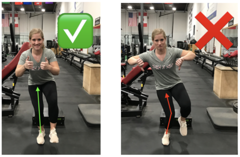
    For example- a patient referred to the clinic with low back pain required to be tested for single leg stance as a part of the examination. The following instructions were given to conduct the test and when one the impairments was identified, that movement was corrected using the same movement as an exercise with specific instructions


    INSTRUCTIONS TO PERFORM THE TEST:- Perform variation if box is checked
    ·       Stand with both feet relatively close together to keep from shifting to the side of the stance leg.

    ·       Shift your weight to stance leg

    ·       Tighten your buttock muscle on the side of your stance leg

    ·       Lift your alternate thigh in front of your body while bending your knee

    o Contract your abdominal muscles
    o Keep your pelvis level ++
    o Place your hands on your pelvis to monitor your movement ++
    o Do not let your opposite hip drop (Figure B)
    o Do not let your knee turn inward (Figure C)
    o Do not let your shoulders lean to the side (Figure D) ++
    keep your trunk still ++
    o Do not let your ankle pronate (i.e., arch collapsed or turned in) ++


    The patient had 2 impairments:- the hip of the stance leg dropped and the knee moved inwards, suggesting a particular impairment which was corrected using cues.

    CONCLUSION:-

    The MSI based classification and treatment allows physical therapists to diagnosis and treat musculoskeletal conditions based on principles of the KPM where impaired alignments and movements are proposed to induce pain and pathology.

    MSI syndromes and treatment have been described for all body regions. The reliability and validity of the system for some anatomical regions have been partially described.85256575859606271727375767778

    Several case reports describing MSI examination and treatment of different musculoskeletal conditions have been published7980818687888990 although efficacy of treatment has not been tested in randomized controlled trials, except in people with chronic low back pain.84

    REFERENCES:-

    Diagnosis and treatment of movement system impairment syndromes

    Shirley Sahrmann, Daniel C. Azevedo, and Linda Van Dillen

    https://www.ncbi.nlm.nih.gov/pmc/articles/PMC5693453/#


  • Prevention Exercises and Tips For ACL Injury


    acl tear
    acl tear

    As Indians are adopting to more sporting lifestyle to maintain fitness or for leisure, the number of ACL tears reported has risen drastically. Earlier the age groups were either sporting population, females or accidental ACL Tears but, recently young adolescents playing sports like Football, Kabaddi, Cricket, etc have incurred ACL tears along with the existing categories.

    Despite thousands of research articles and investigations, the fact that we can predict an ACL injury is only situational…..yes I’m not going to say biomechanics and other factors like all articles. Why situational…..let me enlighten the perspective as per my understanding.

    First of all most Indians who incur an ACL tear, are playing for fun…so when you are playing, the question is that…..is an exercise science genie watching you and telling “hey u landed with a faulty stance, bowl correctly”….if yes then u may be saved, but if not…u might end up with an ACL tear and sit in an orthopods clinic hearing” get reconstruction done otherwise you will get osteoarthritis”…..boom……there goes a gun on your knee…..

    coming back to the prevention topic…normally a physio prehab program must go on for 8-10 weeks for logical and scientific effects. In my experience, I had a chance to conduct a prehab for the under 19 women’s cricket team in Mumbai…although it was just 4 weeks, the results were fantastic. similarly, I had a chance to conduct a musculoskeletal assessment on the Senior Indian Basketball women’s team, upon which, one player was identified to be predisposed to an ACL injury, for which I had implemented a prehab program, which gave fantastic results.

    Having said that. a traumatic ACL tear is not a possibility to prevent unless u gauge/ foresee the injury happening and prevent yourself.

    check out our client assessment done for a post-op ACL tear client ready to enter his 3rd phase of rehab and closer to playing football again

    The prevention program is based on certain principles which we follow at physiocure. If u are a budding athlete or fitness freak, it is a must to learn a program to strengthen the legs to prevent these injuries, and even before that, it is important to identify the population which could be susceptible to ACL tears strategically.

    THE SIX PRINCIPLES OF PREVENTION STRATEGIES (Nessler et all 2017)(1)

    Age

    It is recommended that ACL prevention programs be implemented at an early age. Fewer ACL injuries were documented in younger athletes who performed a neuromuscular training program compared to older athletes who performed the same program

    Figure 7

    Physiocure Exercise Prevention (PEP) programs are based on the above principles. to learn a proper landing technique or jumping technique, we follow the PEP program wherein the biomechanical fault is first examined, and then the required solution is set up as a prehab program for the player.

    for eg:- A basketball player, tall had a habit of playing quickly, but was not able to make quick turns, had a history of knee injuries for which treatment was done. on examination, the jump analysis showed that the player had a habit of letting the knees collapse, which in turn shows poor hip control. Accordingly, a glute activation and strengthening program were recommended along with correct jump landing drills.

    Biomechanics

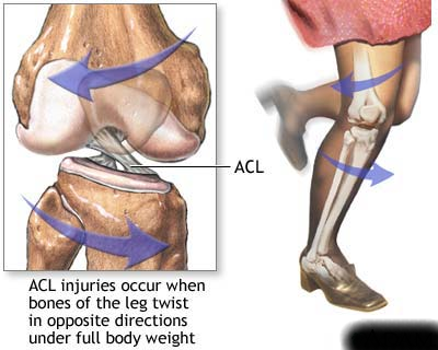
    Faulty biomechanics correlated with increased strain on the ACL during different movement patterns and sporting activities with increased knee valgus being one of the strongest indicators of increased ACL injury risk…..what the last statement means is that when you are landing from a jump, the fact that your knee falls in is one of the biggest predictors that you might get an ACL tear

    Compliance

    Compliance with the performance of an ACL prevention program is vital to the ability of the program to be successful at reducing injury rates. Compliance greater than 66% resulted in an ACL injury reduction rate of 82%. However, when the compliance rate dropped to less than 66%, the ACL injury reduction rate dropped to 44%. Currently, compliance is subject to a large degree of heterogeneity in literature, and the need to have a uniform definition is needed.

    Dosage

    Frequent participation in an ACL prevention program decreases the risk of ACL injury. Most studies agree that each session should be between 20 and 30 min and should be performed several times per week. Optimally, they should be initiated in the pre-season and continued throughout the season to attain the full effectiveness of the program.

    Feedback

    Many studies included some type of feedback, and whether verbal or visual, a decrease in ACL injuries has been shown. Feedback can be in the form of verbal cueing from a coach or a training partner, but can also be visual as in a training video. Feedback should also come in the form of an external focus versus an internal focus. External focus is directed toward the outcome or effects of the movement, assists the automation of movements, and accelerates the learning process. When teaching proper landing mechanics, an external focus command would be, “Try to bring your knees as close to the outside walls as possible when you land”. Internal focus is directed toward specific movements, such as “keep your knees out,” and this constant focus on correct movement can lead to a reduction in athlete’s motivation. Thus, it is recommended that a feedback system be implemented in an ACL prevention program with an external focus.

    Exercise Variety

    ACL injury prevention programs that included a variety of different exercises have a greater incidence of decreasing injury risk versus programs that include only one type of exercise or component (plyometric, balance, strengthening). These exercises can be mainly classified into three different components: plyometrics, neuromuscular training, and strength training. All three have been utilized as stand-alone programs or have been combined to create comprehensive prevention programs.

    check out these 10 simple exercises to improve balance

  • Why Do You Get ACL Injuries

    Sometimes knowing just why do you get ACL injuries can help u clear your mind and take the next step well. The reason I came up with a separate article on why do ACL injuries occur is that I’ve met more than 100 clients who experienced confusion, mind fogging or anxiety post their injury and were not able to decide what next step to take.

    obviously when you incur the injury at first pain and swelling and disability make thinking a little difficult and trust me not know what to do can delay the treatment options.

    Another reason why one must know why do these injuries occur so frequently is for the prevention of ACL injuries. Today, science has given us data, which helps us predict what all factors can lead to ACL injuries. using this information, physios design a prehab program for athletes who are prone to ACL injuries and help saving healthcare costs for that particular team.

    <

    FACTORS INCREASING SUSCEPTIBILITY TO ACL INJURIES:- 

    Gender bias:-

    Female athletes have been reported to sustain non-contact ACL injuries at a rate two- to eightfold greater than their male counterparts. Many explanations for the increased risk of injury to female athletes have been proposed, including increased knee valgus or abduction moments, generalized joint laxity, knee recurvatum, ACL size, and the hormonal effects of estrogen on the ACL

    Improper force generation of quadriceps and hamstrings:-

    when the two thigh muscles namely; Hamstrings and Quadriceps contract together, they create a protective compression force on the knee joint which helps prevent ACL injuries. research has identified that lack of this force generation has lead to ACL tears.

    Frontal Plane Motion


    (more…)