Currently a very small % of women exercise at the recommended levels in India. In fact most obstetricians recommended avoiding exercise.
There are lot of myths in terms of exercising during pregnancy, namely
- Physical activity has detrimental effects such as increasing body temperature.
- There is an increased risk of congenital abnormalities.
- Shifting oxygenated blood & energy away from the developing foetus, thus leading to disturbance in growth of the baby.
All these and many more and no scientific research or proof to prove them true.
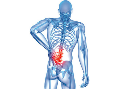
Usually a normal pregnancy lasts for 36-40 weeks in which the woman gains around 10-15 kgs. The common physical problems associated with pregnancy are cramps, varicose veins, symphysis pubis dysfunction, carpal tunnel syndrome, rib pain, stress incontinence, tiredness & Low Back Pain
Anatomical and biomechanical changes during pregnancy
It is important to understand certain anatomical regions of the bones and muscles supporting the pregnant uterus as well their effects on our daily activities.
The Pelvis
The pelvic girdle is an important anatomical structure with respect to pregnancy. Relaxation of the pelvis is essential during pregnancy in order to make room for the growing fetus . The anatomy of the pelvic girdle as follows:
- The pelvic ring includes the sacrum, the sacroiliac joints, the ilium, pubis and ischium and the pubic symphysis
- Unlike the male pelvis which is narrow, deep and oblong the female pelvis is wide, shallow, roomy and round. The main differences are that the subpubic angle is wider and the inlet plane is bigger in the female pelvis.
- In the pelvis the sacroiliac joints are the connection of the spine and legs
- The sacro iliac joints (SIJ) are primarily stabilized by the anterior sacroiliac ligament, interosseous ligament and short and long posterior sacroiliac ligaments
- Muscles that stabilize the SIJ joint are erector spinea, lumbar multifidi, abdominal muscle group (external and internal obliques, rectus abdominis and transverus abdominis) and hamstrings
When the female pelvis does not conform to the normal alignment problems can occur. A non-optimal relation between the maternal pelvis and the fetal head may lead to complications during childbirth, potentially necessitating a caesarean section (Wilder 1988). Maternal causes include earlier pelvic trauma and innate malformations. Fetal causes include hydrocephalus disturbed circulation of cerebrospinal fluid which leads to brain dilation and cranial expansion.
Pelvic floor muscles
- The pelvic floor muscles are important during the pregnancy and the delivery.
- The pelvic diaphragm consists of the levator ani and coccygeus muscles which support the pelvic viscera.
- The coccygeus muscle in addition, flexes the coccyx. The pelvic wall consists of the piriformis and obturator internus muscles which also have a function at the hip joint.
- The pelvic floor consists of the sphincter and erector muscles.
Abdominal wall muscles
- The anterior abdominal wall consists of the rectus abdominis muscle which acts to flex the trunk.
- There are internal and external oblique muscles which act together to bend the trunk to the same side and rotate to the opposite side.
- The obliques have a bilateral function to flex the trunk, compress abdomen and stabilize the pelvis.
- These muscles are directed vertically, horizontally and oblique.
- During pregnancy the diameter of the abdomen increases due to which some women develop a lateral separation of the rectus abdominus, which may persist even after the pregnancy.
- The function of these muscles seems to be the same as with non-pregnant woman.
- The ability of the abdominal muscles to stabilize the pelvis against resistance has been shown to be less in the third trimester of the pregnancy and this stays with the majority of the women until at least 8 weeks after birth.
Biomechanics
- During pregnancy the total body mass increases as the fetus develops, which increase the body weight and changes the physical loading.
- As the gravid uterus increases in size, the centre of gravity shifts forward, causes the pelvis to move in the front and may result in an increased curvature in the lower back and anterior flexion in the neck .
- Vertical forces applied to the feet during dynamic activities, such as walking, running and jumping, reach larger magnitudes ranging from two to five times body weight. However the majority of the mass is retained in the trunk region.
- Getting up from a chair while pregnant causes a 33% increase in the tibiofemoral joint force, an 83% increase in patella femoral force, 100% increase in the activity of the quadriceps muscles and 35% increase in activity in the hamstring muscles.
http://physiocure.in/updates/pregnancy-and-exercises/safety-issues-in-pregnancy/
http://physiocure.in/updates/pregnancy-and-exercises/effects-of-exercises-in-pregnancy/
[contact-form][contact-field label=”Name” type=”name” required=”1″][contact-field label=”Email” type=”email” required=”1″][contact-field label=”Message” type=”textarea”][/contact-form]